University of Kentucky Chandler Medical Center Lexington, KY
Award: ACG Presidential Poster Award
Rebecca Aquino, MD1, Kshitij Thakur, MD2 1University of Kentucky Chandler Medical Center, Lexington, KY; 2University of Kentucky, Lexington, KY Introduction: Endovascular embolization is a key intervention for ectopic varices, but coil migration into the biliary tree is a rare and underrecognized complication. We present a case of progressive coil erosion into the common bile duct (CBD), initially manifesting as obstructive jaundice and evolving to require serial cholangioscopy-guided coil debulking and stenting.
Case Description/
Methods: A 37-year-old man with cryptogenic cirrhosis and prior transjugular intrahepatic portosystemic shunt (TIPS) placement presented with painless jaundice and marked hyperbilirubinemia. He had a remote history of isolated duodenal varix treated with endovascular coiling. Initial endoscopic retrograde cholangiopancreatography (ERCP) revealed a distal CBD stricture with coil material adjacent to the duct. A plastic biliary stent was placed.
On follow-up ERCP, balloon sweeps caused visible movement of the adjacent coil, raising suspicion for intraductal erosion. Cholangioscopy with a single-use digital cholangioscope confirmed erosion of coil fragments into the bile duct lumen. Targeted removal of coil fragments was performed via the major papilla. Biliary stents were replaced to maintain ductal patency. The patient’s bilirubin levels normalized, though repeat ERCPs have been required for ongoing coil fragment debulking. Discussion: Biliary obstruction secondary to coil migration is an extremely rare delayed complication of variceal embolization. This case illustrates a dynamic progression from unexplained biliary stricture to confirmed coil erosion. Serial ERCPs combined with cholangioscopy provided both diagnostic clarity and therapeutic control.
Single-use cholangioscopy was crucial for safe, direct visualization and extraction of embedded coil fragments. Clinicians should consider coil migration in patients with prior embolization who present with biliary strictures or cholestasis and recognize that management may require a staged, multidisciplinary approach.
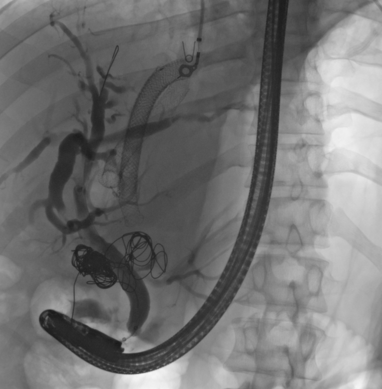
Figure: Cholangiogram showing duodenal coils causing external compression on the mid CBD.
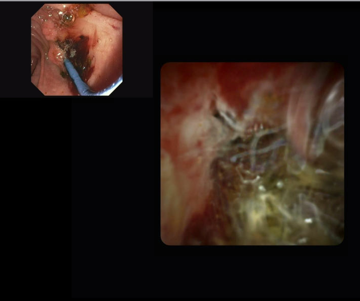
Figure: Cholangioscopy image showing eroded coil in the bile duct.
Disclosures: Rebecca Aquino indicated no relevant financial relationships. Kshitij Thakur indicated no relevant financial relationships.
Rebecca Aquino, MD1, Kshitij Thakur, MD2. P3621 - Uncoiled: Endovascular Coil Erosion into the Bile Duct Causing Obstructive Jaundice and Managed with Serial Cholangioscopy-Guided Debulking, ACG 2025 Annual Scientific Meeting Abstracts. Phoenix, AZ: American College of Gastroenterology.