Monday Poster Session
Category: Interventional Endoscopy
P3590 - A Minimally Invasive Approach to Hepatic Sarcoidosis: Diagnosis via Endoscopic Ultrasound-Guided Fine-Needle Biopsy
Monday, October 27, 2025
10:30 AM - 4:00 PM PDT
Location: Exhibit Hall

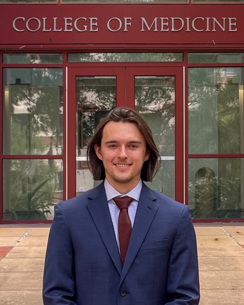
Colin Welch, MS
University of Florida College of Medicine
Gainesville, FL
Presenting Author(s)
Colin Welch, MS1, Zixian Sha, BS1, Lindsey A. Creech, DO, MBA, MPH2, Aleksey Novikov, MD1
1University of Florida College of Medicine, Gainesville, FL; 2University of Florida, Gainesville, FL
Introduction: Sarcoidosis is a multi-system inflammatory disease characterized by non-caseating granulomas. Typical manifestation is intrathoracic; however, a subset of patients has prominent hepatic involvement. There is no current consensus on a diagnostic tool for liver biopsy for patients with or suspected of sarcoidosis. We present two patients diagnosed with hepatic sarcoidosis using endoscopic ultrasound-guided fine needle biopsy (EUS-FNB).
Case Description/
Methods: Case 1
A 57-year-old man presented with 10 days of jaundice, pruritus, and choluria with elevated LFTs (ALT 184 IU/L, AST 184 IU/L, ALP 185 IU/L). Hepatitis panel, HIV and CMV were negative. RUQ US and CT Abdomen Pelvis indicated hepatic steatosis and prominent nonspecific periportal lymphadenopathy, including a small node in the pancreatic head. EUS with FNB was pursued, and lymph nodes were sampled transduodenally with the 22-gauge (22G) US biopsy needle. Color Doppler imaging was then utilized to confirm a lack of significant vascular structures in the liver, followed by two passes (x2) with the 22G US biopsy needle using a transgastric approach (Left Lobe) and transduodenal approach (Right Lobe). Pathology revealed multiple non-necrotizing granulomata, concerning for granulomatous hepatitis. Subsequent chest CT showed hilar and mediastinal adenopathy, confirming a diagnosis of sarcoidosis. The patient started prednisone with improvement of LFTs within a few months.
Case 2
A 41-year-old woman with pulmonary, sinonasal, and ocular sarcoidosis presents with acute RUQ pain and arthralgias with an elevated LFTs (ALT 38 IU/L, AST 58 IU/L, ALP 594 IU/L). MR abdomen and liver elastography noted prominent abdominal lymph nodes and stage 1 fibrosis. Due to lack of chronic suppressive therapy, hepatic involvement was suspected. Using EUS with FNB, liver tissue sampled using transgastric (x2) and transduodenal approaches (x1) with 19G Acquire biopsy needle. Pathology revealed discrete portal and lobular non-necrotizing epithelioid granuloma confirming hepatic sarcoidosis. Patient was treated with prednisone and adalimumab with improvement in LFTs over the following months.
Discussion: Our cases highlight the safety and efficacy of EUS-FNB for liver biopsy in those with or suspected of hepatic sarcoidosis. EUS-FNB can successfully provide both lymph node and liver biopsies despite using a small needle size, displaying its usefulness in multisystem disease diagnosis and adequate specimen size for tissue acquisition.
Disclosures:
Colin Welch indicated no relevant financial relationships.
Zixian Sha indicated no relevant financial relationships.
Lindsey Creech indicated no relevant financial relationships.
Aleksey Novikov indicated no relevant financial relationships.
Colin Welch, MS1, Zixian Sha, BS1, Lindsey A. Creech, DO, MBA, MPH2, Aleksey Novikov, MD1. P3590 - A Minimally Invasive Approach to Hepatic Sarcoidosis: Diagnosis via Endoscopic Ultrasound-Guided Fine-Needle Biopsy, ACG 2025 Annual Scientific Meeting Abstracts. Phoenix, AZ: American College of Gastroenterology.
