Ian Tobal, DO1, Sameera Shuaibi, MD2, Jennifer von Ende, MD1, Paula Cacioppo, MD1, Mohammad Sultan, DO2, Reinaldo Quevedo, MD1 1Ochsner Medical Center, New Orleans, LA; 2Ochsner Health Clinic Foundation, New Orleans, LA Introduction: Hemosuccus pancreaticus (HP) is a hemorrhage from the ampulla of Vater passing through the main pancreatic duct toward the duodenum. Presenting signs and symptoms include epigastric pain that radiates to the back, melena, and hematochezia. Severe cases may present with hematemesis and shock. The abdominal pain may be undulating with fluctuating rates of bleeding, secondary to the movement of the pancreatic ducts. This diagnosis is associated with alcohol use and chronic pancreatitis. HP is a challenging diagnosis to make given its rarity, anatomic location, and ability to mimic other diagnoses such as PUD or gastritis. It carries a mortality risk of 9.6% but with supportive care only, this number approaches 90%.
Case Description/
Methods: A 69-year-old male with a history of alcoholic cirrhosis and esophageal varices presented with four days of melena, severe, undulating epigastric pain, and hypotension. Shortly after admission, he also began to have hematemesis. Lab work was notable for a Hgb of 4.4, a lipase of 402, and a lactic acid of 2.6. He promptly underwent EGD with push enteroscopy that showed blood in the second portion of the duodenum and brisk bleeding from the ampulla of Vater. IR was consulted to perform an angiogram and found a large pseudoaneurysm arising from a branch of the superior pancreaticoduodenal artery. The pseudoaneurysm was embolized with control of the bleeding and resolution of abdominal pain. Discussion: It is estimated that 1 in 1,500 cases of GIB are secondary HP. Causes include chronic pancreatitis, arterial aneurysms, pancreatic tumors or pseudocysts, and iatrogenic insults. This case shows several unique aspects in the diagnosis of HP. Firstly, patients do not commonly present with hematemesis, and when they do, it is important to promptly stabilize them. Also, we were fortunate to see active bleeding from the Ampulla of Vater with a forward-facing endoscope, which is seen in only in 30% of cases. Both components guided the rapid diagnosis and treatment of a life-threatening bleed. Other considerations in the diagnosis of HP include the use of ultrasound, which can help identify abdominal aneurysms or pancreatic pseudocysts in a time or resource-limited setting. Angiography remains the optimal approach as it provides both diagnostic and therapeutic modalities. While IR embolization is the most common intervention and is often successful, there remains a 30% chance of re-bleeding after treatment.
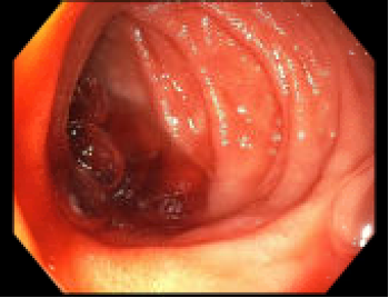
Figure: EGD image showing active bleeding from the ampulla of Vater with blood pooling in the duodenum
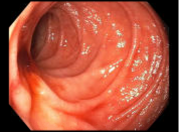
Figure: EGD showing active bleeding from the ampulla of Vater
Disclosures: Ian Tobal indicated no relevant financial relationships. Sameera Shuaibi indicated no relevant financial relationships. Jennifer von Ende indicated no relevant financial relationships. Paula Cacioppo indicated no relevant financial relationships. Mohammad Sultan indicated no relevant financial relationships. Reinaldo Quevedo indicated no relevant financial relationships.
Ian Tobal, DO1, Sameera Shuaibi, MD2, Jennifer von Ende, MD1, Paula Cacioppo, MD1, Mohammad Sultan, DO2, Reinaldo Quevedo, MD1. P3092 - Hemosuccus Pancreaticus: An Unusual Source of GI Bleeding, ACG 2025 Annual Scientific Meeting Abstracts. Phoenix, AZ: American College of Gastroenterology.