Pei-Hsuan Li, DO, Melissa Hershman, MD, BSN Oregon Health & Science University, Portland, OR Introduction: Appendiceal mucocele (AM) is a rare condition characterized by mucin accumulation causing progressive dilatation the appendiceal lumen. It is often discovered incidentally on ultrasound or CT imaging and may mimic acute appendicitis. Endoscopically, AM may appear as a smooth, submucosal bulge at the appendiceal orifice or as mucin secretion into the cecum. While many cases are benign, AM carries malignant potential, particularly in patients with risk factors such as a personal or family history of colorectal cancer, female gender, younger age, or inflammatory bowel disease. We present a case of AM incidentally discovered during colonoscopy without correlating findings on CT imaging.
Case Description/
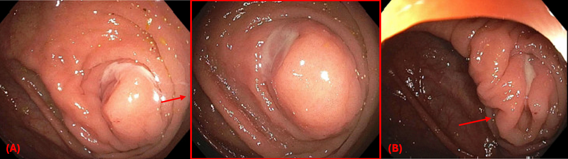
Methods: A 43-year-old female presented for a follow up colonoscopy 3 months following acute uncomplicated sigmoid diverticulitis diagnosed by CT imaging. Family history was pertinent for a gastrointestinal malignancy, subtype unknown, in her mother. The endoscopic exam was normal, apart from mucin secretion from a bulging appendiceal orifice, raising suspicion for AM (Figure 1). The patient was referred for surgical evaluation. However, repeat CT showed a normal appendix, and laboratory testing including tumor markers (CEA, CA19-9, CA 125) was unremarkable. She underwent a laparoscopy and appendectomy, with no suggestion of peritoneal disease and normal appearing appendix. Final pathology demonstrated fibrofatty obliteration of the distal lumen and benign lymph nodes, with no evidence of mucinous epithelial neoplasm. The patient recovered well postoperatively. Discussion: AM represents a spectrum of pathology ranging from simple retention cysts to mucinous adenocarcinomas, with malignancy accounting for up to 20% of cases. Although typically detected on imaging, AM may lack radiographic evidence. This case underscores the diagnostic importance of recognizing the classic endoscopic features, which present as mucin secretion or a smooth bulging lesion at the appendiceal orifice. Timely diagnosis and surgical intervention are crucial to mitigate the risk of rupture and peritoneal dissemination, especially in patients with elevated cancer risk. Even when imaging appears normal as in this case, a high index of suspicion based on endoscopic findings should prompt surgical evaluation to rule out malignancy.
Figure: Figure 1: Endoscopy demonstrated a bulging appendiceal orifice (A) with mucin visibly exuding from the lumen (B)
Disclosures: Pei-Hsuan Li indicated no relevant financial relationships. Melissa Hershman indicated no relevant financial relationships.
Pei-Hsuan Li, DO, Melissa Hershman, MD, BSN. P3016 - Mucin on Scope, Nothing on Scan: Endoscopic Diagnosis of Appendiceal Mucocele in the Absence of Imaging Findings, ACG 2025 Annual Scientific Meeting Abstracts. Phoenix, AZ: American College of Gastroenterology.

 photo")