James E. McSweeney, MD1, Jeff Anucha, MD1, Katherine Waters, BS2, Nathaniel A. Kirchoff, BS3 1South Georgia Medical Center, Valdosta, GA; 2South Georgia Medical Center, Macon, GA; 3South Georgia Medical Center, Savannah, GA Introduction: Dysphagia lusoria is a rare vascular cause of dysphagia stemming from vascular compression of the esophagus by an aberrant subclavian artery¹. The aberrant artery most commonly arises from the left-sided aortic arch passing posterior to the esophagus and causing compression. Several mechanisms have been proposed to explain the onset of symptoms such as increased rigidity of esophageal or vascular walls with aging, aneurysm formation with high-risk features like Kommerell’s diverticulum, age-related elongation of aorta and presence of other anomalies like a bicarotid trunk². Treatment options range from dietary modifications to surgical intervention in more severe cases.
Case Description/
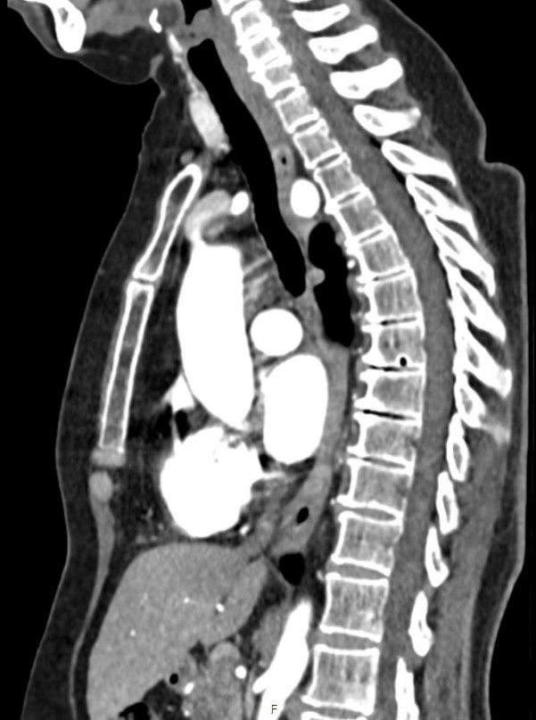
Methods: A 64 year old woman presented with intermittent pharyngeal dysphagia and choking episodes. She reported no associated weight loss. Fluoroscopic barium swallow was largely unrevealing. Upper endoscopy was negative for any gross lesions but did show narrowing of the upper esophagus with concern for questionable external compression (Fig. 1). CTA of the chest confirmed an aberrant retroesophageal right subclavian artery causing mild anterior displacement of the esophagus (Fig. 2). She was referred to vascular surgery and underwent right subclavian-carotid bypass with thoracic endovascular aortic repair stenting. Following surgery she reported notable improvement in swallowing. Postoperative recovery was uneventful and the patient was discharged home with follow-up recommendations including dietary modifications and routine monitoring. Discussion: Although dysphagia is a common clinical complaint encountered in daily practice this case highlights the importance of thorough evaluation to identify rare causes like dysphagia lusoria. Appropriate investigation including fluoroscopic barium swallow, upper endoscopy and advanced imaging with computed tomography ultimately led to the identification of an aberrant right subclavian artery compressing the esophagus.
References:
1. Polguj, M., Chrzanowski, Ł., Kasprzak, J. D., Stefańczyk, L., Topol, M., & Majos, A. (2014). The Aberrant Right Subclavian Artery (Arteria Lusoria): The Morphological and Clinical Aspects of One of the Most Important Variations—A Systematic Study of 141 Reports. The Scientific World Journal, 2014, 292734.
2. Fukuhara, S., Patton, B., Yun, J., & Bernik, T. (2013). A novel method for the treatment of dysphagia lusoria due to aberrant right subclavian artery. Interactive Cardiovascular and Thoracic Surgery, 16(3), 408–410.
Figure: Fig. 1 External compression of the middle third of the esophagus by aberrant subclavian artery.
Figure: Fig. 2 Right subclavian artery is noted to coarse posterior to the esophagus. There is mild anterior displacement of the esophagus by the aberrant subclavian artery.
Disclosures: James McSweeney indicated no relevant financial relationships. Jeff Anucha indicated no relevant financial relationships. Katherine Waters indicated no relevant financial relationships. Nathaniel Kirchoff indicated no relevant financial relationships.
James E. McSweeney, MD1, Jeff Anucha, MD1, Katherine Waters, BS2, Nathaniel A. Kirchoff, BS3. P2870 - Aberrant Right Subclavian Artery and Dysphagia Lusoria, ACG 2025 Annual Scientific Meeting Abstracts. Phoenix, AZ: American College of Gastroenterology.