Kyle Schneider, MD1, Chelsea Wuthnow, MD, JD2, Paul Tarnasky, MD2 1Methodist Dallas Medical Center, Arlington, TX; 2Methodist Dallas Medical Center, Dallas, TX Introduction: Esophageal adenocarcinoma (EA) is a relatively rare diagnosis in the United States. Typical symptoms of EA are dysphagia, chest pain, cough, and weight loss. While metastases to liver, lung and lymph nodes can often be the presenting symptoms, metastasis to the spinal column is exceedingly rare with only a few cases documented. Here, we report a case of metastatic cancer that was incidentally discovered during spinal decompression surgery that was due to an undiagnosed EA.
Case Description/
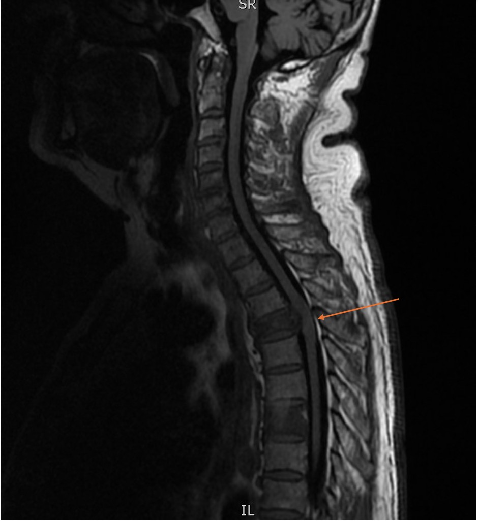
Methods: A 67-year-old male presented with chief complaint of chest pain and leg weakness. He was found to have pulmonary embolism with incidental osseous metastasis at the level of T3-T4 (Image 1). Follow up imaging demonstrated thoracic cord compression and he was taken for surgery where a fungating mass was found. Surgical pathology revealed a CK7+, CK20-, CDX2+ adenocarcinoma most consistent with upper gastrointestinal or pancreatobiliary primary. Prior to admission, he endorsed some early satiety but denied any other upper gastrointestinal symptoms including dysphagia, reflux, and/or melena. No other sites of metastases were noted. Esophagogastroduodenoscopy (EGD) showed long segment Barrett’s esophagus in the middle and lower third of the esophagus along with severe mucosal changes characterized by erythema, friability, and nodularity, along with one non-bleeding gastric ulcer (image 2). Esophageal biopsy revealed Her2-, microsatellite stable, PDL1+ esophageal adenocarcinoma. Discussion: Metastasis from a primary EA is rare, and metastatic disease to the spine is even more rare. In this case and in 3 other reported cases, spinal metastasis is an indicator of poor prognosis. Typical presenting symptoms include dysphagia, early satiety, and odynophagia. Risk factors for development of EA can include long-standing reflux along with Barrett’s esophagus. In this case, the patient did not have complaints of dysphagia. EGD can be beneficial for diagnosis of primary EA. If neurological symptoms develop and persist, magnetic resonance imaging with gadolinium should be included. This case highlights both the rarity of metastatic spinal EA and the need for a multidisciplinary approach as well as shared decision making with the patient. EA may present with distant metastases in the absence of classic gastrointestinal symptoms, underscoring the need for a broad differential when evaluating spinal lesions of unknown origin.
Figure: Image 1: MRI Image of metastatic lesion to T4 (spinal compression noted by arrow)
Figure: Image 2: EGD imaging of lower third of esophagus with long Barrett's and mucosal changes
Disclosures: Kyle Schneider indicated no relevant financial relationships. Chelsea Wuthnow indicated no relevant financial relationships. Paul Tarnasky indicated no relevant financial relationships.
Kyle Schneider, MD1, Chelsea Wuthnow, MD, JD2, Paul Tarnasky, MD2. P2862 - A Real Pain in the Neck - A Rare Presentation of Esophageal Adenocarcinoma, ACG 2025 Annual Scientific Meeting Abstracts. Phoenix, AZ: American College of Gastroenterology.