Baton Rouge General Medical Center Baton Rogue, LA
Rineetha Tandra, MD, MBBS1, Nagendra Babu Indurti, MD2, Ogochukwu Ugochukwu, MBBS3, Chimdalu C. Ezeani, 4, Chukwunonso Ezeani, MBBS5, Andrew Nelson, MD6 1Baton Rouge General Medical Center, Baton Rogue, LA; 2Baton Rouge General, Baton Rouge, LA; 3University of Nigeria, Baton Rouge, LA; 4University of Nigeria College of Medicine, Ituku-Ozalla, Enugu, Nigeria; 5Mayo Clinic, Phoenix, AZ; 6Gastroenterology Associates, Baton Rouge, LA Introduction: Esophageal submucosal hematoma is an uncommon condition caused by rupture of submucosal blood vessels. Most cases present with chest pain, vomiting, or dysphagia soon after the inciting cause. Only a small proportion of cases present following a fall in elderly patients. We report the case of a 76-year-old lady who experienced sudden onset neck pain and dysphagia after falling at home while taking apixaban, which mimicked esophageal cancer on imaging.
Case Description/
Methods: A 76-year-old woman presented to the emergency room with bilateral neck pain and difficulty swallowing. She fell out of her bed onto a carpeted floor around 2 AM while having a nightmare. She did not lose consciousness. Her medical history includes chronic atrial fibrillation on Eliquis and breast cancer status lumpectomy and radiation.
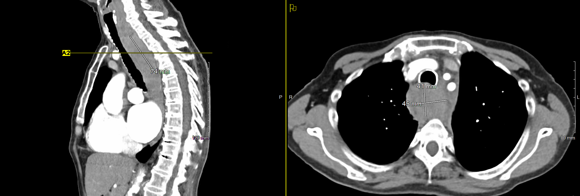
On presentation, she was tachypneic, but other vital signs were stable. Her physical exam was unremarkable. The complete blood count was normal. Non-contrasted computed tomography of her cervical spine revealed prominent soft tissue thickening in the cervical and upper esophageal/retrotracheal soft tissue. Possible esophageal wall thickening with intraluminal debris was also noted. Based on the recommendation of the radiologist, a follow-up contrast-enhanced CT chest was done and showed a 4.1 x 4.5 cm marked esophageal prominence with near-complete luminal occlusion in the superior thoracic esophagus. This finding was suspicious for malignancy (figure 1).
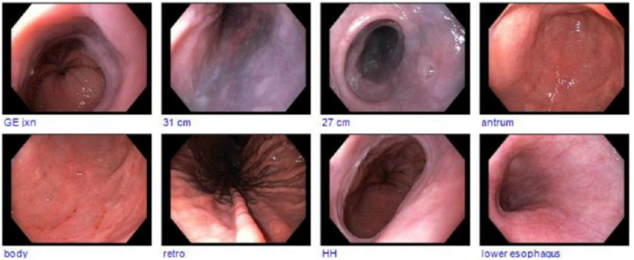
She was admitted, placed on IV fluids, NPO, pantoprazole, and apixaban was held. The gastroenterology team was consulted, and the patient underwent esophagoduodenoscopy (EGD) on day 2, which revealed semicircumferential purple discoloration from 27 to 31 cm from the incisors, consistent with a submucosal hematoma. There was no ulcer or mass seen (figure 2). Dysphagia resolved during hospitalization. She was discharged with instructions to hold apixaban for two days. On follow-up few months later, patient's symptoms have completely resolved. Discussion: We have presented the case of a 76-year-old lady on apixaban who presented with dysphagia following a fall at home. This case highlights that esophageal submucosal hematoma can result from minor indirect injuries, and CT scans may not be diagnostic. Physicians should have a high index of suspicion to differentiate hematoma from cancer, especially in the absence of weight loss.
Figure: Figure 1: CT Chest with IV contrast showing esophageal prominence
Figure: Figure 2: EGD showing submucosal hematoma
Disclosures: Rineetha Tandra indicated no relevant financial relationships. Nagendra Babu Indurti indicated no relevant financial relationships. Ogochukwu Ugochukwu indicated no relevant financial relationships. Chimdalu Ezeani indicated no relevant financial relationships. Chukwunonso Ezeani indicated no relevant financial relationships. Andrew Nelson indicated no relevant financial relationships.
Rineetha Tandra, MD, MBBS1, Nagendra Babu Indurti, MD2, Ogochukwu Ugochukwu, MBBS3, Chimdalu C. Ezeani, 4, Chukwunonso Ezeani, MBBS5, Andrew Nelson, MD6. P2849 - Esophageal Hematoma: An Uncommon Case of Acute Dysphagia, ACG 2025 Annual Scientific Meeting Abstracts. Phoenix, AZ: American College of Gastroenterology.