Mark McGarrey, MD, Harrison Pajovich, MD, Stefanie Gallagher, DO, MBe, Lauren Davis, DO, Erin Hollis, DO, Michelle Springer, DO Lankenau Medical Center, Wynnewood, PA Introduction: Appendiceal neuroendocrine tumors (NETs) are rare, usually asymptomaticand often discovered incidentally during appendectomy for presumed appendicitis. We present a case of a patient presentingwith acute, recurrent abdominal pain and imaging suggestive of early sigmoid volvulus. She had a normal-appearing sigmoidoscopy but had continued pain and a concerning abdominal exam.An exploratory laparoscopy was performed which revealed an inflamed appendixand she underwent an appendectomy with pathology revealing aneuroendocrine microadenoma.
Case Description/
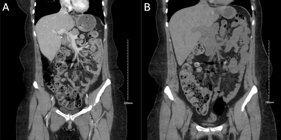
Methods: A 30-year-old woman with history of irritable bowel syndrome and irondeficiency anemia presentedwith acute onset right lower quadrant (RLQ) abdominal pain, nausea,vomiting, and constipation. Labs notable for hemoglobin of 11.6 g/dL, white bloodcell count of 7.8, and lactate of 0.5 mmol/L. CT abdomen and pelvis showed minimallydilated sigmoid colon displaced to the right abdomen with mild narrowing and surroundingfat stranding concerning for early sigmoid volvulus or internal hernia involving the sigmoidcolon (Figure 1A). Chart review revealed 3 similar presentations prior to this with unrevealing cross-sectional imaging and work up.The patient was taken for flexible sigmoidoscopy which showed redundant andtortuous sigmoid colon without narrowing, stricture, or malrotation. Repeat CT of theabdomen and pelvis showed no evidence of sigmoid volvulus (Figure 1B). The patient continued tohave worseningRLQ pain with rebound tendernessand guarding. She was taken for exploratory laparoscopy which showedan inflamedappendix, normal appearing sigmoid colon and scattered pelvic endometriosis. Pathological analysis of the excised appendixrevealed a 0.5 mm neuroendocrine microadenoma with Ki-67 proliferation index of ~1%. Her pain resolved post-operatively. Discussion: Appendiceal NETs are the most common neoplasm of the appendix,found in approximately 0.3–0.9% of appendectomy specimens, most often in young adultsand are often discovered incidentally during surgery for acute appendicitis. This case highlights a rare instance of appendiceal NET causing intermittent obstruction only to be diagnosed once acute appendicitis developed.Imaging concerning for sigmoid volvulus, a more common cause of intermittent abdominal pain that classically presents with sigmoid distension, tapering of the bowel lumen, and twisting of the mesenteric vessels on CT imaging, acted as a red herring and added to the diagnostic complexity of this case.
Figure: Coronal CT images of the abdomen before and after flexible sigmoidoscopy. (1A) Distended sigmoid colon displaced toward the right abdomen with tapering suggestive of early sigmoid volvulus. (1B) Interval normalization of sigmoid configuration following flexible sigmoidoscopy, consistent with a redundant but non-obstructed sigmoid colon.
Disclosures: Mark McGarrey indicated no relevant financial relationships. Harrison Pajovich indicated no relevant financial relationships. Stefanie Gallagher indicated no relevant financial relationships. Lauren Davis indicated no relevant financial relationships. Erin Hollis indicated no relevant financial relationships. Michelle Springer indicated no relevant financial relationships.
Mark McGarrey, MD, Harrison Pajovich, MD, Stefanie Gallagher, DO, MBe, Lauren Davis, DO, Erin Hollis, DO, Michelle Springer, DO. P2587 - Recurrent Abdominal Pain With a Twist: An Unexpected Appendiceal NET, ACG 2025 Annual Scientific Meeting Abstracts. Phoenix, AZ: American College of Gastroenterology.

 photo")