The Warren Alpert Medical School of Brown University Warwick, RI
Gurjot Singh, DO1, Tamara Koritarov, DO, MSc2, Cristina Hamacher, DO, PharmD3, Shevonne Marcelle, DO4, Elizabeth Decker, DO5 1The Warren Alpert Medical School of Brown University, Warwick, RI; 2Kent Hospital, Providence, RI; 3Brown University/Kent Hospital, Warwick, RI; 4Warren Alpert Medical School of Brown University, Warwick, RI; 5Kent Hospital, Warwick, RI Introduction: Desmoplastic small round cell tumors (DSRCTs) are a very rare type of sarcoma with a poor prognosis. Around 200 cases are said to be reported in literature and even more rarely in the rectum. Here we present a case of a DSRCT with a primary site originating from the rectum.
Case Description/
Methods: A 29-year-old male with no past medical history presented with bright red blood per rectum (BRBPR) for 3 months. He reported 15-pound unintentional weight loss, lower abdominal discomfort, occasional night sweats, rare alcohol use, with no family history of any major gastrointestinal pathologies. On admission, complete blood count was found to be unremarkable with a hemoglobin of 14.9 g/dL, white blood cell count, and platelets within normal limits. Iron level was found to be low at 39 mcg/dL, a TIBC of 272 mg/dL, transferrin of 218 mg/dL, and ferritin of 79.4 ng/dL. Hepatitis panel, HIV testing, and CEA were unremarkable. CT scan of the chest, abdomen, and pelvis revealed a large soft tissue mass measuring 10.8 x 8.9 x 9.5 cm circumferentially encasing the rectum with surrounding lymphadenopathy notable within the left mesorectal fat and the aortic bifurcation insistent of metastatic disease. No evidence of obstruction noted. MRI of the abdomen completed demonstrated a 2 cm and 1.1 cm lesion of the liver suspicious for metastatic disease. Colonoscopy revealed a partially obstructed mass from 5 to 22 cm proximal to the anus biopsied with initial pathology demonstrating small cell carcinoma. Left rectal lymph node biopsy was consistent with initial diagnosis of small cell carcinoma. PET scan demonstrated FDG avid rectal mass, lymphadenopathy at aortic bifurcation, left external iliac chain, two liver lesions, and left ischial lesion. Rectal biopsy was sent for genetic testing with EWSR1: WT1 fusion with RNA sequencing and found to have a final diagnosis of desmoplastic small round cell tumor. The patient was started on vincristine, doxorubicin, and cyclophosphamide therapy. Discussion: Desmoplastic small round cell tumors is a sarcoma which affects primarily young males and is associated with a unique chromosomal translocation of chromosome 11 and 12 leading to oncoprotein formation of EWSR1:WT1. Very few numbers of cases have been reported in literature with primary sites of the abdomen and pelvis causing symptoms of abdominal distension. Less than approximately 5 cases have been reported with the primary site of the rectum with an unusual presentation of rectal bleeding.
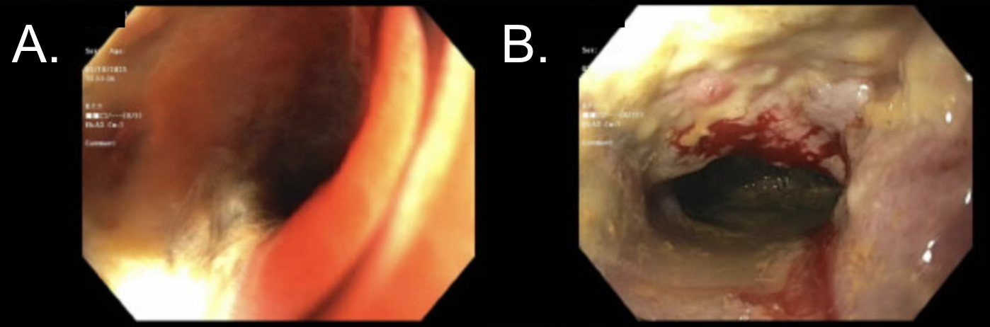
Figure: Colonoscopic findings of (A.) mass found at the sigmoid colon, 20 cm from the anal verge (B.) mass continuing throughout the sigmoid colon 15 cm from the anal verge.
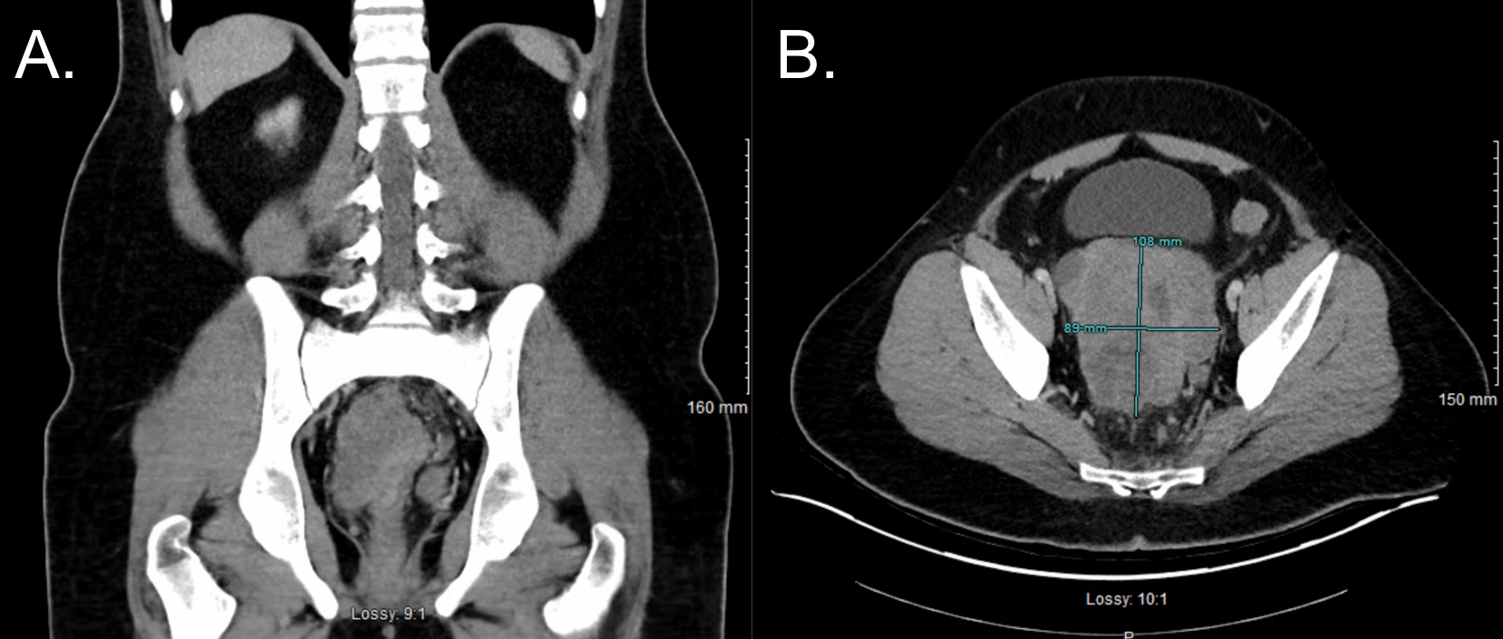
Figure: CT findings of the abdomen and pelvis demonstrating rectal mass (A.) coronal view (B.) axial view with rectal mass measured to be 10.8 x 8.9 cm.
Disclosures: Gurjot Singh indicated no relevant financial relationships. Tamara Koritarov indicated no relevant financial relationships. Cristina Hamacher indicated no relevant financial relationships. Shevonne Marcelle indicated no relevant financial relationships. Elizabeth Decker indicated no relevant financial relationships.
Gurjot Singh, DO1, Tamara Koritarov, DO, MSc2, Cristina Hamacher, DO, PharmD3, Shevonne Marcelle, DO4, Elizabeth Decker, DO5. P2528 - A Very Rare Case of Desmoplastic Small-Round-Cell Tumor With Primary Rectal Involvement in a 29 Year-old Man, ACG 2025 Annual Scientific Meeting Abstracts. Phoenix, AZ: American College of Gastroenterology.