University of Hawaii, John A. Burns School of Medicine, Department of Medicine Honolulu, HI
Award: ACG Presidential Poster Award
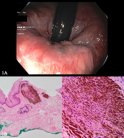
Arvin Jeremy N. Tan, MD1, Akiko Tokunaga, MD2, Toru Nakata, MD, PhD1, Yusuke Miyatani, MD1, Vishal Dobaria, MD1, Chad Cryer, MD3, Traci T. Murakami, MD, FACG4 1University of Hawaii, John A. Burns School of Medicine, Department of Medicine, Honolulu, HI; 2University of Hawaii, John A. Burns School of Medicine, Department of Pathology, Honolulu, HI; 3University of Hawaii, John A. Burns School of Medicine, Department of Surgery, Honolulu, HI; 4The Queen's Medical Center, Ewa Beach, HI Introduction: Melanocytic nevi are small, round, uniformly colored, benign clonal proliferation of pigment-producing cells. They are typically found in the skin, particularly on sun-exposed areas. Its occurrence in the rectum is rare, with an undetermined incidence and prevalence. Here, we present a case of an asymptomatic 55-year-old female who was found to have a melanocytic nevus of the rectum.
Case Description/
Methods: A 55-year-old female presented to the gastroenterology clinic for a screening colonoscopy. She was asymptomatic without a family history of colorectal malignancy or colonic polyps. She has not had prior endoscopies. During the procedure, a small, raised, hyperpigmented lesion was detected on the rectum below the dentate line upon retroflexion view (Image 1A). The lesion was not biopsied at that time. The patient was referred to surgery and subsequently underwent complete surgical excision of the lesion. Pathologic examination revealed a submucosal melanocytic nevus without malignant transformation or extension to the margins (Image 1B-1C). She tolerated the procedure well and was scheduled for follow-up colonoscopy after 10 years. Discussion: Melanocytic nevi of the rectum are rare, with an estimated prevalence of 0.21%. While benign, they have the potential for malignant transformation into malignant melanoma. Less than 1% of all cases of malignant melanoma occur in the anorectal area, however, it accounts for up to 4% of all anorectal malignancies with a poor prognosis. Early detection of precursor lesions like melanocytic nevi is therefore important. There is currently no established standard of treatment for rectal melanocytic nevi. The current practice stems from a strong consideration of the risk of malignant transformation. Several options include surveillance with periodic colonoscopy, and endoscopic or surgical removal. This case highlights the importance of vigilance for non-polypoid lesions during endoscopy and of doing a careful retroflexion examination for the detection of lesions that may otherwise be missed.
Figure: Image 1A: Colonoscopy image in retroflexion view of a small, raised, hyperpigmented lesion on the rectum. 1B-1C: Photomicrograph of the lesion demonstrating a nest of melanocytes in the dermis without cytologic atypia.
Disclosures: Arvin Jeremy Tan indicated no relevant financial relationships. Akiko Tokunaga indicated no relevant financial relationships. Toru Nakata indicated no relevant financial relationships. Yusuke Miyatani indicated no relevant financial relationships. Vishal Dobaria indicated no relevant financial relationships. Chad Cryer indicated no relevant financial relationships. Traci Murakami indicated no relevant financial relationships.
Arvin Jeremy N. Tan, MD1, Akiko Tokunaga, MD2, Toru Nakata, MD, PhD1, Yusuke Miyatani, MD1, Vishal Dobaria, MD1, Chad Cryer, MD3, Traci T. Murakami, MD, FACG4. P2518 - Beyond the Skin: A Case of Melanocytic Nevus of the Rectum, ACG 2025 Annual Scientific Meeting Abstracts. Phoenix, AZ: American College of Gastroenterology.