
Monday Poster Session
Category: Colon

Nisar Amin, MD
Charleston Area Medical Center
Charleston, WV
Liposarcoma of the colon is exceptionally rare, as most cases arise in the retroperitoneum or extremities. Despite its rarity, recognizing its clinical features, diagnostic challenges, and treatment strategies is crucial. We report a case of colonic liposarcoma presenting as a large intraluminal mass.
Case Description/
Methods:
A 40-year-old male with long-standing history of colitis presented with lower abdominal pain, frequent bowel movements, maroon-colored stools. Family history was notable for multiple malignancies, including ovarian, bone, skin, and small-cell cancers.
On exam, he was hemodynamically stable with mild diffuse abdominal tenderness. Laboratory findings included microcytic anemia (Hg 10.7 g/dL, MCV 78 fL) and an iron profile consistent with iron deficiency anemia. Abdominal CT scan revealed a large, 14.5 x 16.1 x 22 cm fatty retroperitoneal mass arising near the right adrenal region with mass effect and calcified soft tissue components. Tumor markers (CEA and CA 19-9) were normal. Colonoscopy revealed a 5 cm firm, semi-pedunculated mass distal to hepatic flexure (Figures A, B). Biopsies showed acute inflammatory exudate and underlying florid granulation tissue, no malignancy was noted. However, a CT-guided biopsy of retroperitoneal mass revealed leiomyosarcoma. He underwent surgical resection, including a 30 cm segment of right and transverse colon. Pathology demonstrated a dedifferentiated liposarcoma (Grade 3) composed of well-differentiated, smooth muscle/fibrous, and high-grade pleomorphic sarcoma components (Figure C). Tumor involved the submucosa of colon and omentum and extended to radial margin. The patient had an uneventful postoperative recovery and was discharged with plan to follow up with oncology for outpatient consideration of adjuvant chemotherapy.
Discussion:
Liposarcoma of the colon is extremely rare, particularly the dedifferentiated subtype, with only two primary cases reported in PubMed between 1990 and 2016. This subtype is associated with poor prognosis, exhibiting higher rates of recurrence and mortality compared to other variants. Standardized diagnostic and treatment guidelines are limited due to the rarity of cases. Surgical resection is the mainstay of treatment, often followed by adjuvant chemotherapy. While typically seen in the fifth to sixth decades, earlier onset can occur. Nonspecific symptoms and deep tissue involvement often lead to diagnostic challenges and false-negative biopsies, warranting a high index of suspicion in atypical cases.
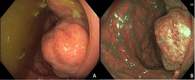
Figure: Figures A (white light exam) and B (narrow band imaging exam) showing a 5 cm firm, semi-pedunculated mass distal to the hepatic flexure.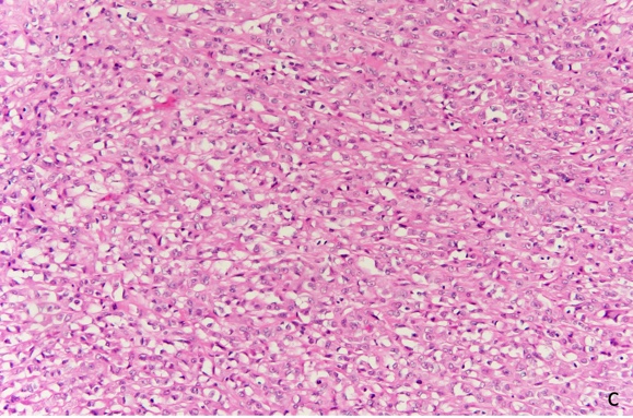
Figure: Figure C: Histopathology showing soft tissue tumor with features of dedifferentiated liposarcoma. The tumor demonstrates hypercellular, high grade round cell to pleomorphic sarcoma. (H&E, 20X).
Disclosures:
Nisar Amin indicated no relevant financial relationships.
Mai Tran indicated no relevant financial relationships.
Ebubekir Daglilar indicated no relevant financial relationships.
Harleen Chela indicated no relevant financial relationships.
Nisar Amin, MD, Mai T. Tran, MD, Ebubekir Daglilar, MD, Harleen Chela, MD. P2516 - Dedifferentiated Liposarcoma Presenting as a Colonic Mass: A Case Report, ACG 2025 Annual Scientific Meeting Abstracts. Phoenix, AZ: American College of Gastroenterology.