
Monday Poster Session
Category: Colon

Sirisha Gaddipati, MD
University of Miami Health System
Miami, FL
Multiple myeloma (MM) is a common plasma cell malignancy; however, extramedullary plasmacytoma (EMP)—plasma cell proliferation outside of bone marrow— is an aggressive and uncommon phenotype. Gastrointestinal (GI) involvement is exceedingly rare. We present a case of multifocal secondary colonic EMP in a patient with relapsed refractory multiple myeloma (RRMM) and review literature of reported GI associated EMP.
Case Description/
Methods:
A 70-year-old man with new onset constipation and bone marrow biopsy-proven MM, who failed multiple lines of chemotherapy, underwent PET imaging which revealed increased focal metabolic activity within the hepatic flexure and sigmoid colon (Figure 1a). Diagnostic colonoscopy showed a partially circumferential, partially obstructing mass in the distal ascending colon involving two-thirds of luminal circumference with a luminal diameter of 10 mm, biopsies were performed. In addition, a 17mm sessile cecal polyp and a 4mm transverse colonic semi-pedunculated polyp were endoscopically resected (Figure 1b). Pathology from all sites demonstrated plasmacytomas, staining positive for CD138 and MUM1, and were kappa restricted (Figure 1c). Subsequent labs revealed bicytopenia and atypical lymphocytes with plasmacytoid morphology, suggestive of plasma cell leukemia. He ultimately began treatment with etoposide, prednisone, vincristine, cyclophosphamide, and doxorubicin (EPOCH) as bridging therapy prior to chimeric antigen receptor (CAR)-T cell infusion for treatment of RRMM and currently remains on CAR-T therapy.
Discussion:
While primary EMPs are more common and arise in the absence of systemic MM, secondary EMPs are rare and are associated with with 15% lower 5-year survival outcomes than those without plasmacytoma at MM diagnosis. Gastrointestinal involvement is extremely rare and endoscopic evaluation is required for definitive diagnosis. GI presentations can include constipation, bowel obstruction, and rectal bleeding. Chemotherapy is typically the mainstay of treatment and surgery is usually avoided unless colonic obstruction is present. In our literature review of secondary EMP cases, we found 11 cases reported to date of isolated GI involvement— extensive and multifocal colonic involvement, as seen in our case, was reported in only 1 other case (Table 1). Extramedullary disease typically portends a higher mortality in progressive MM; therefore, prompt recognition and treatment of colonic EMPs is crucial.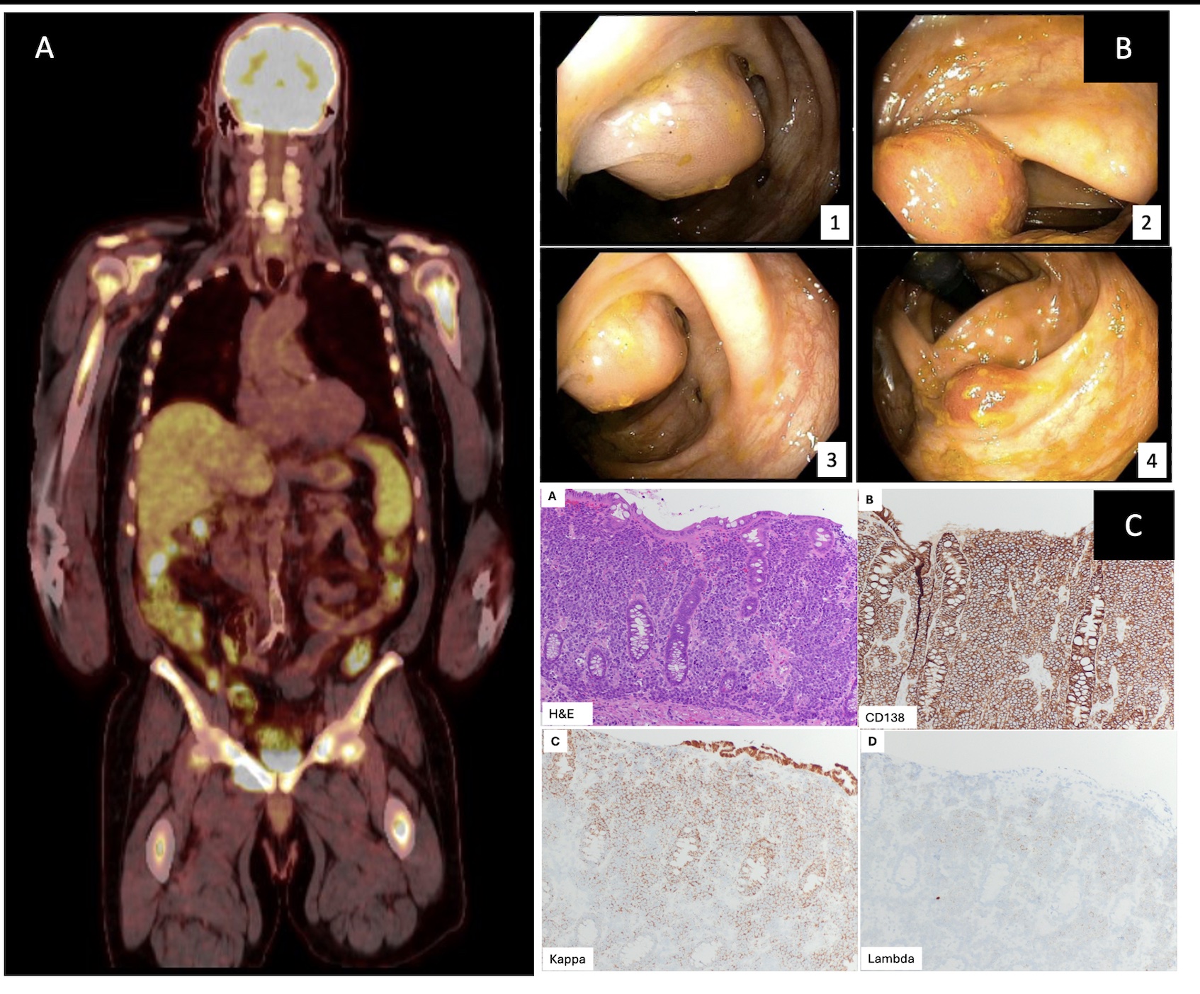
Figure: Figure 1: Radiographic, endoscopic, and histologic evidence of colonic EMP. A) Coronal section of PET scan with focal abnormal metabolic activity noted within the hepatic flexure and sigmoid colon. B) Evidence of polyps on initial colonoscopy including ascending colon (1-3) and cecum (4). C) Histologic findings of ascending colon mass. The large intestinal mucosa is involved by sheets of plasma cells (a) The plasma cells are positive for CD138 (b) and are kappa-restricted by kappa and lambda in situ hybridization stain (c, d).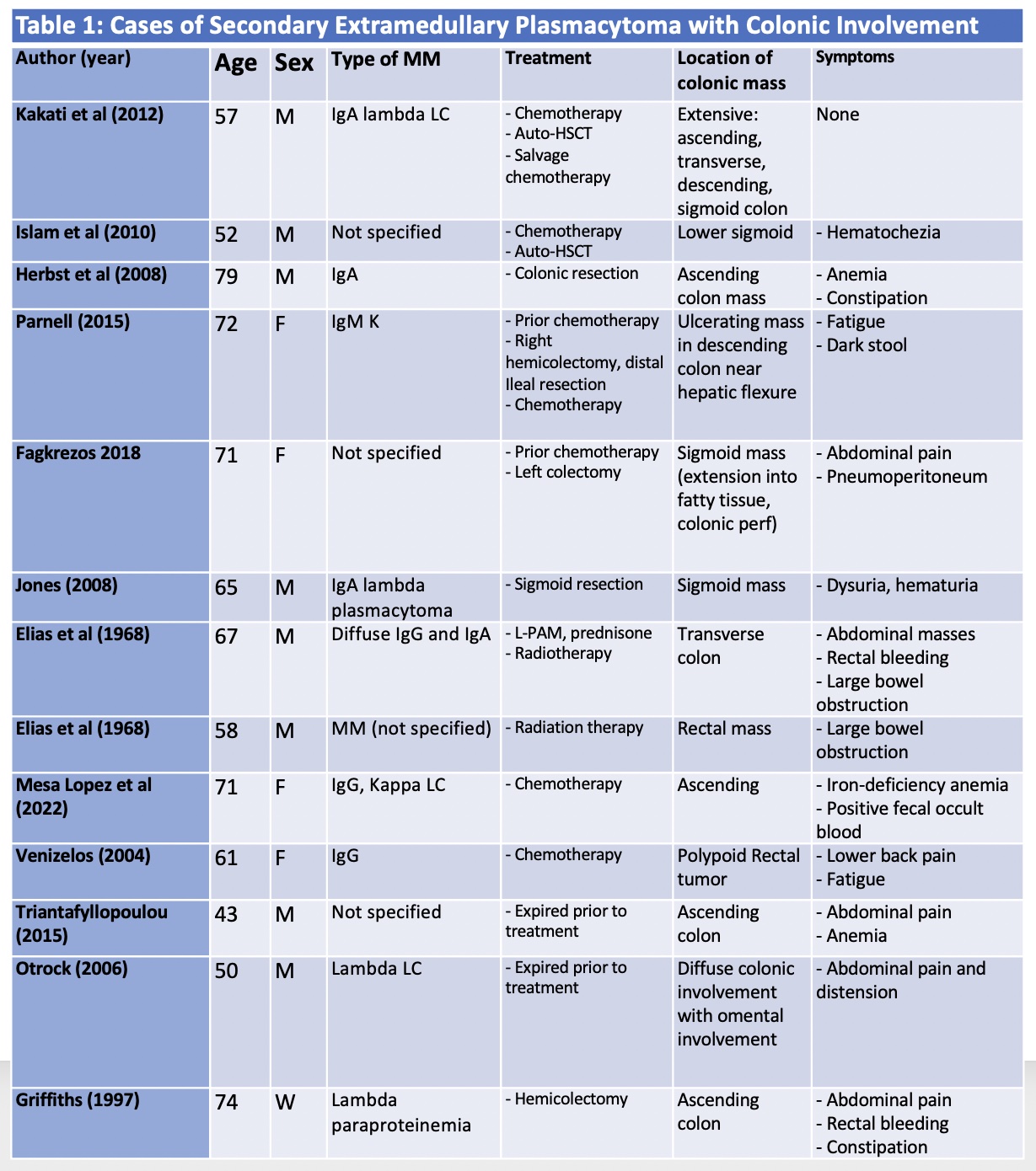
Figure: Table 1: Literature Case Review of Secondary Extramedullary Plasmacytoma with Colonic Involvement.
Disclosures:
Sirisha Gaddipati indicated no relevant financial relationships.
Sunny Sandhu indicated no relevant financial relationships.
Guannan Zhang indicated no relevant financial relationships.
Xiaoqiong Wang indicated no relevant financial relationships.
Eric Martin indicated no relevant financial relationships.
Sirisha Gaddipati, MD1, Sunny Sandhu, MD2, Guannan Zhang, MD1, Xiaoqiong Wang, MD, PhD1, Eric F. Martin, MD3. P2491 - Multifocal Colonic Masses Presenting as Secondary Extramedullary Plasmacytoma in Relapsing-Refractory Multiple Myeloma: A Case Report and Review of Literature, ACG 2025 Annual Scientific Meeting Abstracts. Phoenix, AZ: American College of Gastroenterology.