Ross University School of Medicine cedar falls, IA
Muhammad F. Aftab, MD1, Muhammad Abdullah, MBBS2, Abdullah Javed, MBBS3, Charanjeet Singh, MD4 1Ross University School of Medicine, Miramar, FL; 2Lahore Medical and Dental College, Lahore, Punjab, Pakistan; 3Allama Iqbal Medical College, Lahore, Punjab, Pakistan; 4AdventHealth Orlando, Orlando, FL Introduction: Adenomatoid tumors (ATs) are benign mesothelial neoplasms, rarely arising in the peripancreatic region. When located near the pancreas, these tumors often mimic malignant pancreatic lesions due to their atypical location and imaging characteristics. Herein, we present a case of a peripancreatic AT initially suspected to be a cystic neoplasm.
Case Description/
Methods: A 73-year-old woman with alcoholic chronic pancreatitis and fatty atrophy of the pancreas, presented with a two year history of progressive upper abdominal pain and bloating. An initial MRI demonstrated a 1.5 x 1.2 cm lesion in the left upper quadrant, in close proximity to the body of the pancreas. Further evaluation with endoscopic ultrasound (EUS) revealed a 1.2 cm solid-cystic mass in the body/tail of the pancreas, suspicious for Intraductal Papillary Mucinous Neoplasm (IPMN) or Neuroendocrine Tumor (NET), prompting fine-needle aspiration (FNA). However, the biopsy revealed a proliferation of oncocytic cells without any high grade cytologic features and immunohistochemistry (IHC) showed positivity for cytokeratin AE1/AE3, Cytokeratin 7, WT-1, Calretinin and a low MIB-1 index. It was diagnosed as a benign mesothelial tumor. After discussion in the tumor board, it was decided that no immediate intervention was needed. Serial abdominal imaging showed a stable appearance and EUS was repeated in two years for re-evaluation of the lesion. The findings were consistent with a peri-pancreatic mass, separate from the pancreas and a repeat biopsy showed adenomatoid tumor. It was decided that further surveillance or intervention will be directed by oncology. Discussion: ATs predominantly arise in the genital tract. Extragenital growth of AT in peripancreatic tissue is exceedingly rare with only limited reports. They pose a significant diagnostic challenge, as they may resemble solid-cystic neoplasms of pancreas. Histologically, these tumors are characterized by well-circumscribed lesions. IHC staining is crucial for accurate diagnosis, typically showing positivity for mesothelial markers including, WT1, calretinin, and cytokeratins. This case illustrates that AT exhibits indolent behavior and should be considered in the differentials of atypical peripancreatic lesions. Stable disease on serial imaging and benign histopathology permitted conservative management, sparing unnecessary intervention.
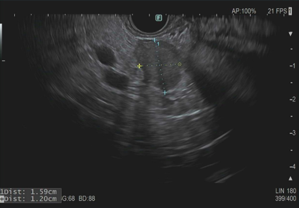
Figure: Figure 1. Endoscopic ultrasound (EUS) showing a well-circumscribed, mixed echogenicity peripancreatic mass with solid and cystic components, measuring 1.59 × 1.20 cm, separate from the pancreas and spleen. EUS-guided fine-needle biopsy was performed.
Disclosures: Muhammad Aftab indicated no relevant financial relationships. Muhammad Abdullah indicated no relevant financial relationships. Abdullah Javed indicated no relevant financial relationships. Charanjeet Singh indicated no relevant financial relationships.
Muhammad F. Aftab, MD1, Muhammad Abdullah, MBBS2, Abdullah Javed, MBBS3, Charanjeet Singh, MD4. P2315 - The Peripancreatic Masquerader: Adenomatoid Tumor Mimicking Common Pancreatic Lesions, ACG 2025 Annual Scientific Meeting Abstracts. Phoenix, AZ: American College of Gastroenterology.

 photo")