University of Texas at Austin Dell Medical School Austin, TX
Mahnoor Liaqat, MD1, Priyanka Jagannathan, MD2, Rima Shobar, MD, MS1, Deepak Agrawal, MD, MPH2, David Tang, MD1 1University of Texas at Austin Dell Medical School, Austin, TX; 2University of Texas at Austin, Austin, TX Introduction: Pancreatic acinar cell carcinoma (PACC) is a rare exocrine pancreatic neoplasm accounting for only 1-2% of all adult pancreatic malignancies. Patients present with non-specific symptoms such as weight loss, nausea, abdominal pain, vomiting, and diarrhea. While elevated serum lipase is commonly observed in PACC, elevated alpha-fetoprotein (AFP) is rare and can mislead diagnosis.
Case Description/
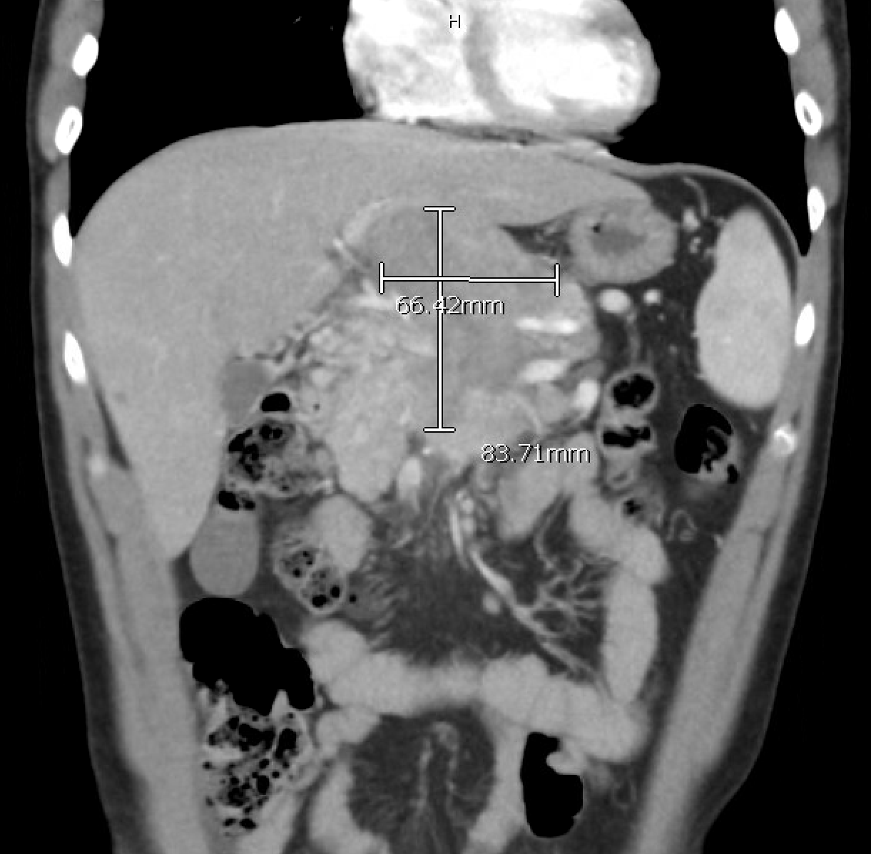
Methods: A 32-year-old male with a history of pre-diabetes presented with chronic worsening right upper quadrant abdominal pain and unintentional weight loss for several months. His physical exam was unremarkable aside from right upper quadrant tenderness. Initial laboratory workup was noted for normal CBC, CMP, lipase levels and elevated LDH (245 units/L). CT abdomen/pelvis with contrast showed a large 5.9 × 6.6 × 8.4cm heterogeneous mass in the pancreatic region with splenic artery encasement and superior mesenteric artery displacement, enlarged para-aortic lymph nodes (23 mm), mild splenomegaly (14.1cm), and a 4mm hepatic lesion. Serum tumor markers showed markedly elevated alpha-fetoprotein ( >20,000 ng/mL), normal CEA and Ca 19.9. Given the elevated AFP there were initial concerns for non-seminomatous testicular germ cell tumor prompting a testicular ultrasound which was normal. Endoscopic ultrasound (EUS) guided fine needle biopsy of the pancreatic mass confirmed acinar cell carcinoma with characteristic uniform acinar morphology, abundant granular eosinophilic cytoplasm, and positive immunohistochemical staining for trypsin and AFP. The tumor showed negative staining for chromogranin, synaptophysin, and hepatocyte markers, confirming acinar differentiation. Discussion: This case represents one of the few reported cases of pancreatic acinar cell carcinoma in a young adult male with extreme AFP elevation ( > 20,000 ng/mL). This case emphasizes the diagnostic perplexity when evaluating patients with a pancreatic mass and markedly elevated AFP highlighting the need to consider this rare entity in the differential diagnosis. Other differentials include pancreatoblastoma, hepatocellular carcinoma, hepatoblastoma and certain lymphomas. Additionally, although the lipase can be significantly elevated in PACC, it may differ by tumor differentiation and a normal lipase does not exclude PACC. Early tissue diagnosis through EUS-guided biopsy is crucial for accurate diagnosis and appropriate management planning.
Figure: AP view of CT scan of mass
Figure: Histology
Disclosures: Mahnoor Liaqat indicated no relevant financial relationships. Priyanka Jagannathan indicated no relevant financial relationships. Rima Shobar indicated no relevant financial relationships. Deepak Agrawal indicated no relevant financial relationships. David Tang indicated no relevant financial relationships.
Mahnoor Liaqat, MD1, Priyanka Jagannathan, MD2, Rima Shobar, MD, MS1, Deepak Agrawal, MD, MPH2, David Tang, MD1. P2255 - When AFP Misleads: Pancreatic Acinar Cell Carcinoma in a Young Adult, ACG 2025 Annual Scientific Meeting Abstracts. Phoenix, AZ: American College of Gastroenterology.