
Tuesday Poster Session
Category: Colon

Anoushka Dua, MD
University of California Irvine Digestive Health Institute
Irvine, CA
Incidental colonic uptake observed on fluorodeoxyglucose positron emission tomography-computed tomography (FDG PET-CT) has been associated with the presence of colonic neoplasms. This study aims to evaluate the diagnostic performance of FDG PET-CT in detecting colonic neoplastic lesions.
Methods:
This IRB-approved retrospective cohort study included all adult patients at a single academic institution who underwent colonoscopy within 12 months of FDG PET-CT between 1/1/2010 and 1/31/2025. Clinical data were extracted from the electronic health record. A PET-CT scan was considered positive if there was any focal FDG uptake in the colon. Colonic neoplasia was defined as the presence of any of the following: hyperplastic polyp, tubular adenoma, sessile serrated adenoma, traditional serrated adenoma, tubulovillous adenoma, any adenoma with high-grade dysplasia, malignant lesions (colorectal origin or metastases), inflammatory polyp, or other polypoid dysplastic lesion.
Descriptive statistics were used to classify PET-CT results as either true positive or false positive for positive scans, and true negative or false negative for negative scans, based on the presence or absence of neoplastic lesions on subsequent colonoscopy in the corresponding region or elsewhere in the colon. These classifications were then used to calculate the sensitivity and specificity of PET-CT for detecting 1) any colonic neoplasm and 2) advanced adenoma or colorectal cancer.
Results:
A query of the electronic health record identified 506 patients who underwent colonoscopy (with adequate bowel preparation) within 12 months of FDG PET-CT. Of these, 304 had negative PET-CT scans and 202 had positive PET-CT scans.
Figure 1 summarizes the classification of neoplastic lesions detected or missed by PET-CT. Figure 2 presents the 2×2 contingency tables used to assess the diagnostic performance of PET-CT for identifying colonic neoplasms. Overall, PET-CT demonstrated a sensitivity of 25.4% and a specificity of 48.2% for the detection of any colonic neoplasm. For advanced adenomas and colorectal cancer, PET-CT showed a sensitivity of 51.2% and a specificity of 61.1%.
Discussion:
While the sensitivity and specificity of FDG PET-CT for detecting advanced precancerous lesions and colorectal cancer remain inferior to those of established non-invasive screening modalities, this study underscores the importance of further evaluation with diagnostic colonoscopy when focal colonic FDG uptake is observed.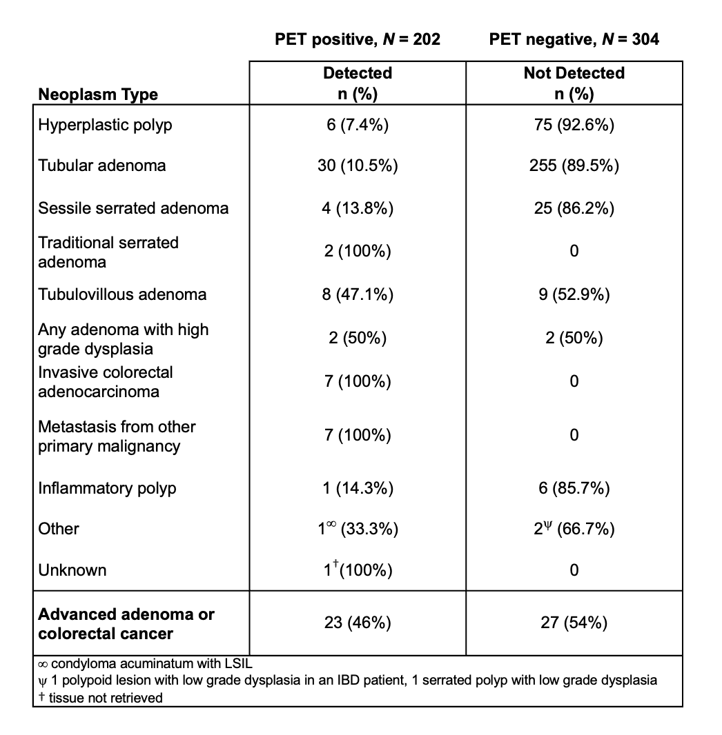
Figure: Summary of colonic neoplastic lesions identified by FDG PET-CT—defined as focal uptake in a colonic region corresponding to a histologically confirmed lesion—and those not detected by FDG PET-CT.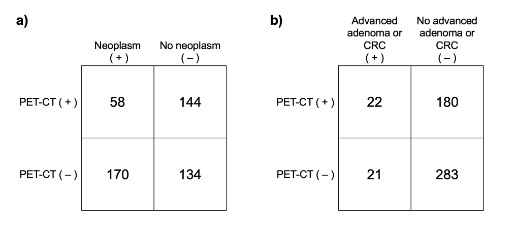
Figure: Contingency tables illustrating the distribution of true positive, false positive, true negative, and false negative PET-CT results, based on colonoscopy findings defined as: (a) any colonic neoplasm, or (b) advanced adenoma or colorectal cancer (CRC).
Disclosures:
Anoushka Dua indicated no relevant financial relationships.
Christian Makar indicated no relevant financial relationships.
Alexander Lee indicated no relevant financial relationships.
Anjali Herekar indicated no relevant financial relationships.
Charlene Yuan indicated no relevant financial relationships.
Valery Vilchez indicated no relevant financial relationships.
William Karnes indicated no relevant financial relationships.
Anoushka Dua, MD1, Christian Makar, MD, MPH2, Alexander Lee, BS2, Anjali Herekar, MD3, Charlene Yuan, MD3, Valery Vilchez, MD3, William Karnes, MD1. P4570 - Sensitivity and Specificity of FDG PET-CT Imaging for the Detection of Colonic Neoplasms, ACG 2025 Annual Scientific Meeting Abstracts. Phoenix, AZ: American College of Gastroenterology.