MedStar Georgetown University Hospital Washington, DC
Usman Afzal, MD1, Thomas Holman, 1, Ahmad Abdulraheem, MD2, Bryce Kunkle, MD1, Ahmad Al-Dwairy, MBBS1, Abdalla Khouqeer, MD1, Walid Chalhoub, MD1 1MedStar Georgetown University Hospital, Washington, DC; 2MedStar Washington Hospital Center-Georgetown University, Washington, DC Introduction: Ganglioneuromas (GN) are rare benign peripheral neuroblastic tumors that arise from neural crest cells. They are usually in the retroperitoneum and the mediastinum; however, pancreatic involvement is rare.
These tumors are usually asymptomatic; however, can present with nonspecific gastroentestinal symptoms due to tumor mass effect. Rarely, ganglioneuromas can also produce vasoactive intestinal polypeptide, androgenic hormones, and catecholamines, leading to flushing, diarrhea, and hypertension. Diagnosis is based on pathology and immunohistochemistry obtained after surgical excision of the tumor.
Case Description/
Methods: We present the case of a 71 year old female who was referred to the gastroenterology clinic for evaluation of an incidental pancreatic cyst seen on computerized tomography (CT) imaging. She was vitally stable and expressed intermittent nausea and 10 pound unintentional weight loss over ten months. She denied smoking, alcohol consumption, illicit drug use, or family history of cancer.
Her lab work was grossly normal, including her complete blood count, basic metabolic panel, liver function tests, bilirubin levels, urinalysis, and TSH. The tumor marker CA 19-9 was 4 U/ml, and IgG subclass 4 was 19 mg/dl.
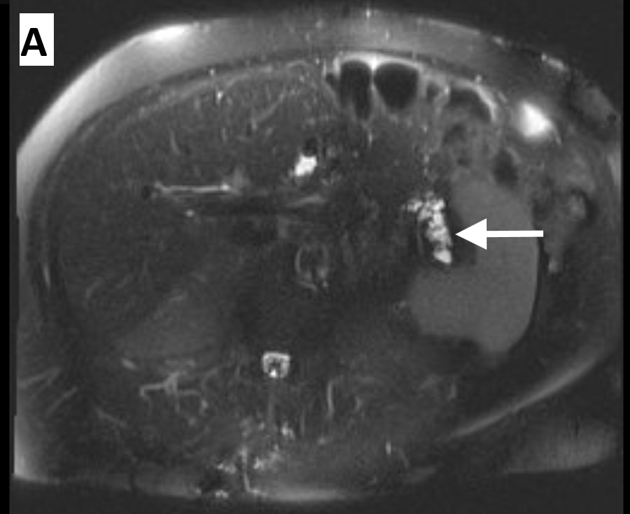
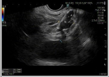
She underwent an MRCP, which showed several cystic lesions of varying sizes throughout the pancreatic parenchyma (Figure 1). She underwent an endoscopic ultrasound (EUS) that showed an ill-defined hypoechoic lesion in the pancreatic tail measuring 26.2 mm x 12.7 mm (Figure 2) that was sampled with fine-needle aspiration and sent for cytology. Pathology was diagnostic of ganglioneuroma. Immunohistochemical staining—positive for S100, SOX10, neurofilament, GFAP, synaptophysin,chromogranin with low Ki-67 proliferation index.
A dotatate positron emission tomography (PET) was obtained that ruled out metastatic disease. Given the benign nature and low malignant potential of the lesion, it was decided to proceed with radiologic surveillance. Discussion: Pancreatic ganglioneuromas are exceedingly rare with less than 10 cases reported in literature. Our case adds to that fund of knowledge since our patient presented at age 71, an outlier from the median age of incidence, 17. It also highlights the importance of comprehensive diagnostic evaluation in patients with pancreatic lesions with tissue sampling since the symptoms at presentation and imaging findings can be indistinguishable between benign ganglioneuromas and ganglioblastomas, which have malignant potential.
Figure: MRCP abdomen showing the pancreatic tail lesions.
Figure: Endoscopic Ultrasound showing an ill-defined hypoechoic lesion in the pancreatic tail measuring 26.2 mm x 12.7 mm
Disclosures: Usman Afzal indicated no relevant financial relationships. Thomas Holman indicated no relevant financial relationships. Ahmad Abdulraheem indicated no relevant financial relationships. Bryce Kunkle indicated no relevant financial relationships. Ahmad Al-Dwairy indicated no relevant financial relationships. Abdalla Khouqeer indicated no relevant financial relationships. Walid Chalhoub indicated no relevant financial relationships.
Usman Afzal, MD1, Thomas Holman, 1, Ahmad Abdulraheem, MD2, Bryce Kunkle, MD1, Ahmad Al-Dwairy, MBBS1, Abdalla Khouqeer, MD1, Walid Chalhoub, MD1. P4462 - Ganglioneuroma of the Pancreas: A Case Report, ACG 2025 Annual Scientific Meeting Abstracts. Phoenix, AZ: American College of Gastroenterology.

 photo")