Department of Internal Medicine, University of Colorado Anschutz Medical Campus, Aurora, Colorado Denver, CO
Abakar Baraka, MD1, Thomas Enke, MD2, Mohammad Bilal, MD, FACG3, Augustin Attwell, MD4 1Department of Internal Medicine, University of Colorado Anschutz Medical Campus, Aurora, Colorado, Denver, CO; 2University of Colorado, Aurora, CO; 3University of Colorado Anschutz Medical Campus, Denver, CO; 4Division of Gastroenterology and Hepatology, Denver Health, Denver, Colorado, Denver, CO Introduction: Congenital malposition of the ampulla of Vater is an exceptionally rare anomaly, often asymptomatic and diagnosed incidentally. However, when clinically significant, it can result in diagnostic confusion and technical difficulty during endoscopy. We present a case of an ectopic ampulla located in the pyloric channel, presenting with biliary obstruction and cholangitis.
Case Description/
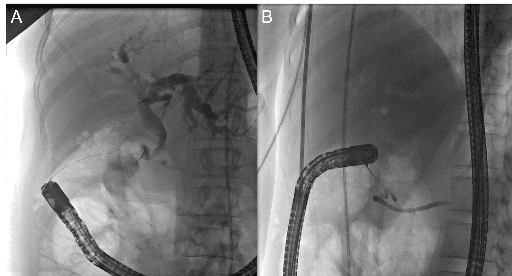
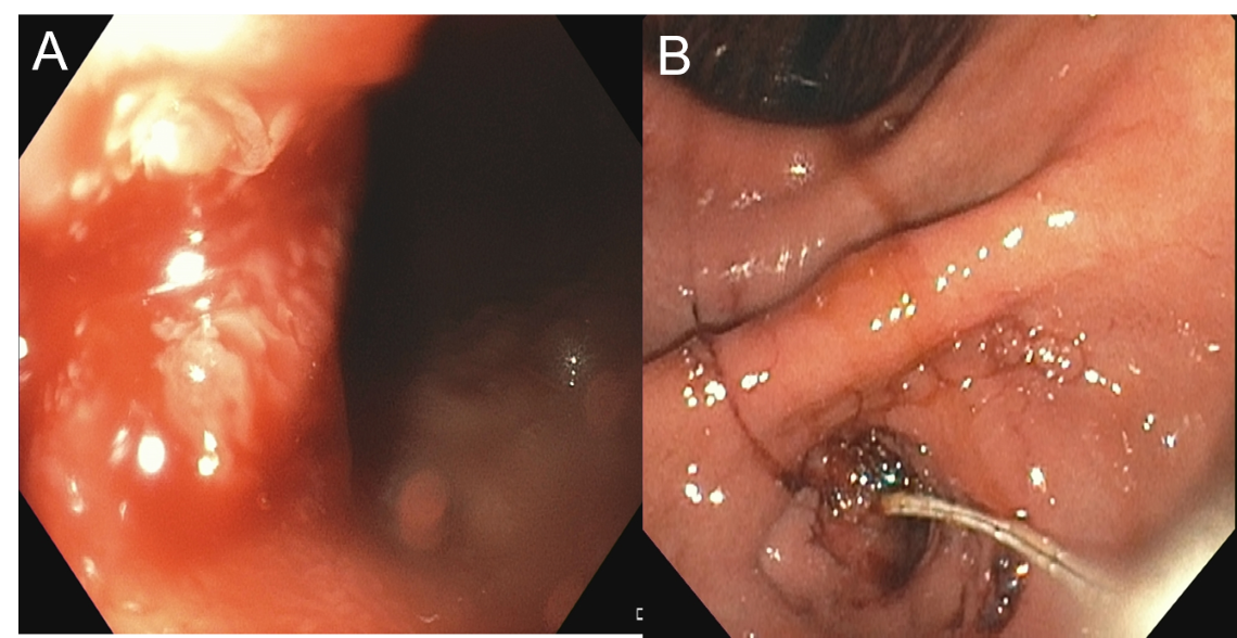
Methods: A 37-year-old woman with a history of remote cholecystectomy due to recurrent cholecystitis presented with epigastric pain, nausea, and mixed liver injury. Notably, magnetic resonance cholangiopancreatography (MRCP) performed a year prior for epigastric pain revealed moderate intra- and extrahepatic biliary dilation with a possible high-grade common bile duct (CBD) stricture. However, further evaluation was deferred due to normal laboratory evaluation and resolution of pain. On current admission, computed tomography of the abdomen re-demonstrated intra- and extrahepatic ductal dilation with suspected CBD stricture and new pneumobilia, raising concern for cholangitis. Endoscopic retrograde cholangiopancreatography (ERCP) was technically challenging due to a severe pyloric stenosis requiring balloon dilation. Despite careful inspection, the major papilla was not identified in the duodenum. Biliary access was ultimately obtained via an ectopic or fistulous opening in the pyloric channel. Biopsies of friable mucosa in the pre-pyloric region were benign. She clinically improved with antibiotics and was discharged. ERCP performed 2 months later more clearly visualized the ampulla of Vater within the pyloric channel (Figure 1) consistent with a congenital ectopic ampulla of Vater. Pancreatogram was normal and cholangiogram revealed a short inflammatory stricture, and distal filling defects (Figure 2). Symptoms resolved with balloon dilation and balloon sweep with removal of several stones and sludge. Discussion: This case highlights a rare congenital anomaly: an ectopic ampulla of Vater located in the pyloric channel. Initially asymptomatic, it led to chronic biliary stasis and ultimately cholangitis due to stone and sludge formation. The diagnosis was delayed despite prior imaging due to normal liver chemistries and lack of symptoms. The case underscores the importance of re-evaluating stable findings in light of evolving clinical changes. Additionally, it illustrates the need for flexibility during ERCP when conventional anatomy is not encountered.
Figure: Figure 1. Ectopic ampulla of Vater located in the pyloric channel (A) and cannulation during endoscopic retrograde cholangiopancreatography (B).
Figure: Figure 2. Final cholangiogram (A) and pancreatogram (B) obtained following cannulation of an ectopic ampulla of Vater located in the pyloric channel.
Disclosures: Abakar Baraka indicated no relevant financial relationships. Thomas Enke indicated no relevant financial relationships. Mohammad Bilal: Boston Scientific – Consultant. Cook endoscopy – Paid speaker. Steris Endoscopy – Consultant. Augustin Attwell indicated no relevant financial relationships.
Abakar Baraka, MD1, Thomas Enke, MD2, Mohammad Bilal, MD, FACG3, Augustin Attwell, MD4. P4440 - An Unexpected Exit: Biliary Obstruction from a Pyloric Ectopic Ampulla, ACG 2025 Annual Scientific Meeting Abstracts. Phoenix, AZ: American College of Gastroenterology.