University of Connecticut Health Center Farmington, CT
Hira Khan, MD1, Karim Al Annan, MD2, Christopher Costa, MD1, Nancy Y. Kang, MD3 1University of Connecticut Health Center, Farmington, CT; 2University of Connecticut Health Center, Hartford, CT; 3Saint Francis Hospital, Trinity Health of New England, Hartford, CT Introduction: Pancreatic pseudocysts (PPCs) are a common complication of pancreatitis, occurring in up to 30% of chronic cases. We present a unique case involving multiple acute and chronic PPC-related complications: intracystic hemorrhage, superior mesenteric vein (SMV) compression, portal vein thrombosis, splenic venous pseudoaneurysm (SVP), and recurrent pancreatitis.
Case Description/
Methods: A 62-year-old man with chronic alcohol-induced pancreatitis and known pancreatic pseudocysts on MRI surveillance presented with weeks of worsening abdominal pain radiating to the back. He was admitted for acute pancreatitis. Vitals showed HR 95 bpm and BP 190/93 mmHg. Exam revealed epigastric tenderness without peritonitis. Labs showed leukocytosis (14.0 x10³/μL), amylase 1397 U/L, lipase 7687 U/L, and ALP 117 U/L.
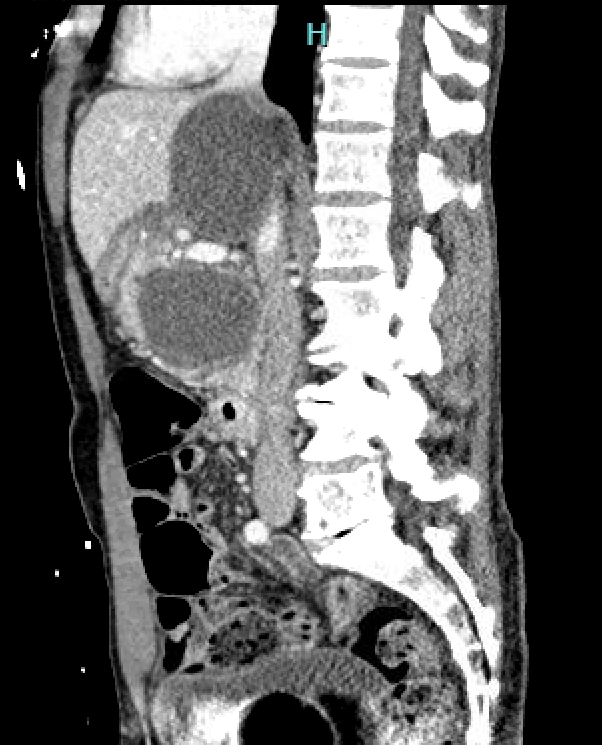
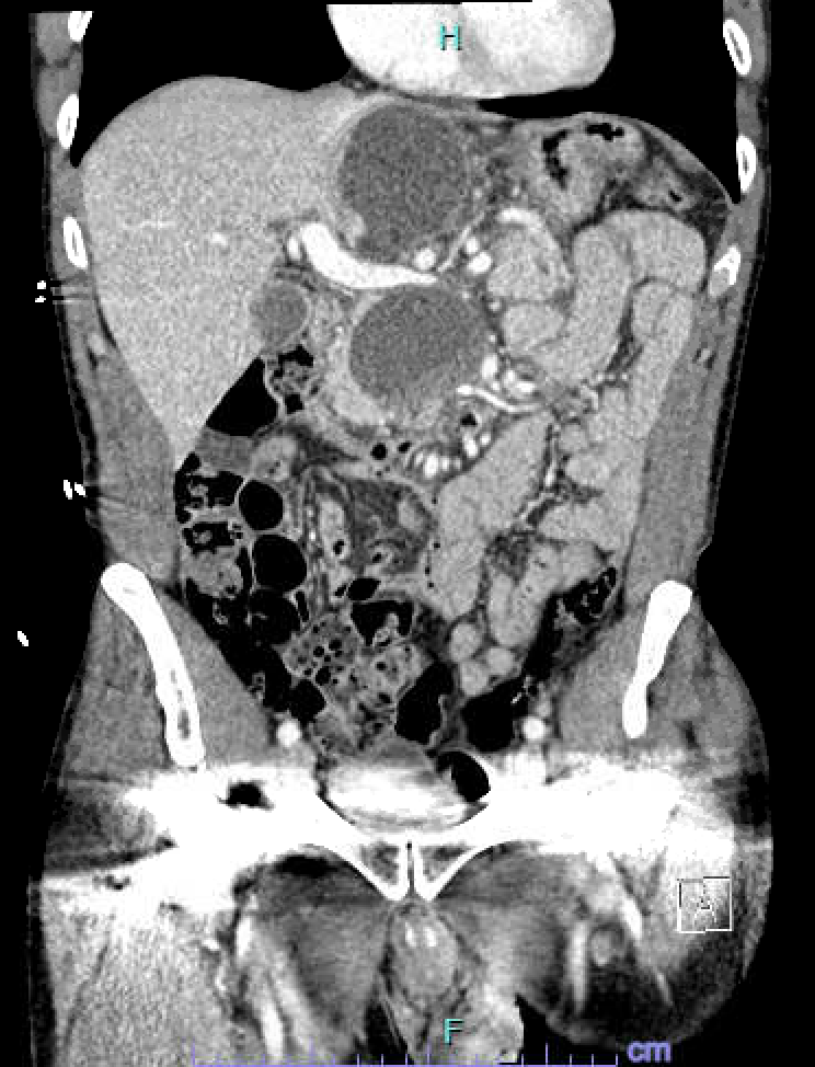
CT abdomen/pelvis revealed two pseudocysts (6.6 cm superior, 5.3 cm inferior) in the uncinate process and pancreatic neck, with peripancreatic fat stranding. The superior PPC caused SMV compression (Figure 1.0); the inferior PPC had increased in size compared to prior imaging and showed new hemorrhage (Figure 2.0). There was also upstream pancreatic duct dilation and thrombosis of the right portal vein branch. CT angiogram revealed splenic vein pseudoaneurysm.
He was managed conservatively with IV fluids and hydromorphone, with symptomatic improvement. He was discharged with plans for outpatient follow-up for somatostatin therapy and subsequent endoscopic drainage of PPCs. Discussion: PPCs may cause hemorrhage, rupture, infection, and compression of adjacent structures including the biliary tree and portal system. This patient had previously undergone endoscopic ultrasound (EUS) aspiration of a 5.3 cm pseudocyst, which showed benign cytology. Five months later, it progressed to 6.6 cm with significant complications.
Although arterial pseudoaneurysms—particularly involving the gastroduodenal artery—are well-documented in association with PPCs, splenic vein pseudoaneurysms remain rarely reported, especially when occurring alongside multiple other complications. This case highlights a complex and unique presentation of a common diagnosis, highlighting the need for close monitoring and consideration of venous complications in patients with PPCs.
Figure: Figure 1.0 showing SMV compression by pseudocysts
Figure: Figure 2.0 showing intra-cystic hemorrhage of inferior pseudocyst
Disclosures: Hira Khan indicated no relevant financial relationships. Karim Al Annan indicated no relevant financial relationships. Christopher Costa indicated no relevant financial relationships. Nancy Kang indicated no relevant financial relationships.
Hira Khan, MD1, Karim Al Annan, MD2, Christopher Costa, MD1, Nancy Y. Kang, MD3. P4399 - When a Pseudocyst Turns Perilous: A Case of Vascular and Pancreatic Complications in Tandem, ACG 2025 Annual Scientific Meeting Abstracts. Phoenix, AZ: American College of Gastroenterology.

 photo")