Detroit Medical Center/Wayne State University Detroit, MI
Kenan Abou Chaer, MD1, Omar Al-Subee, MD, FACG2 1Detroit Medical Center/Wayne State University, Detroit, MI; 2Henry Ford Health, Detroit, MI Introduction: Lung cancer often metastasizes beyond the thorax, commonly to the bones, liver, adrenal glands, regional and distal lymph nodes. Metastasis from lung cancer to the pancreas is rare, and is usually linked to small cell lung carcinoma. Metastasis from non-small cell lung cancer (NSCLC) to the pancreas is particularly uncommon, with one study reporting only 1% incidence. We present a case of biopsy-proven lung adenocarcinoma metastasized into the head of the pancreas with a picture of obstructive jaundice.
Case Description/
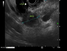
Methods: A 74-year-old female with asthma and presumed lung cancer was treated with Stereotactic Body Radiation Therapy (SBRT) over 7 years due to non-diagnostic biopsies. A PET scan in June 2024 showed diffuse bone metastases and new abdominal lymphadenopathy. A CT-guided iliac bone biopsy confirmed metastatic pulmonary adenocarcinoma (CK7+, TTF-1+, CK20–, CDX2–). She began systemic therapy with pembrolizum, pemetrexed, and carboplatin. In September 2024, she presented with obstructive jaundice and abnormal liver tests (ALP 1156 U/L, AST 234 U/L, ALT 293 U/L, total bilirubin 22.4 mg/dL). CT abdomen showed severe biliary ductal dilation with abrupt tapering at the pancreatic head and multiple liver lesions. A pancreatic head mass was suspected but could not be clearly demonstrated. CA 19-9 was 142 U/mL. ERCP revealed a 2 cm distal CBD stricture, and a 7 cm 10 FR plastic stent was placed. Brushing from distal CBD revealed atypical cells highly suspicious for adenocarcinoma. Subsequently, EUS demonstrated a 23×20 mm hypoechoic pancreatic head mass with poorly defined borders and no vascular invasion. Fine Needle Biopsy (FNB) using a 22-gauge needle on 4 passes confirmed pulmonary adenocarcinoma, matching the bone biopsy stain results. During a second ERCP, a self-expandable partially covered metallic stent was placed. Discussion: Pancreatic metastasis from NSCLC remains a rare but important clinical entity. Differentiating metastatic lesions from primary pancreatic tumors is often difficult due to overlapping clinical and radiographic features. Liver function tests and tumor markers such as CA 19-9 can provide clues to biliary obstruction or metastatic involvement but are nonspecific. Adequate tissue sampling by EUS/FNB with staining remains the ultimate method to confirm such a rare finding, and exclude another coexisting primary adenocarcinoma of pancreas. Distinguishing between primary and metastatic pancreatic tumors is critical, as both treatment and prognosis differ.
Figure: Endoscopic ultrasound (EUS) image showing a 23x20mm pancreatic head mass and a common bile duct (CBD) stent in place.
Disclosures: Kenan Abou Chaer indicated no relevant financial relationships. Omar Al-Subee indicated no relevant financial relationships.
Kenan Abou Chaer, MD1, Omar Al-Subee, MD, FACG2. P4398 - When Lung Cancer Hits the Pancreas: A Case of Rare Metastasis from Non-Small Cell Lung Cancer (NSCLC), ACG 2025 Annual Scientific Meeting Abstracts. Phoenix, AZ: American College of Gastroenterology.