Farah Abuazzam, MD1, Bilind S. Ismail, MD1, Michael Chammany, DO2, Hussien Haidari, MD3, Hassan Khanani, MS4, Sathya Jaganmohan, MD5 1Willis Knighton Health, Shreveport, LA; 2Edward Via College of Osteopathic Medicine, Galveston, TX; 3University of South Alabama, Mobile, AL; 4Edward Via College of Osteopathic Medicine, Shreveport, LA; 5GastroIntestinal Specialists, Shreveport, LA Introduction: Tubulovillous adenoma (TVA) is a benign epithelial neoplasm typically found in the colon and rectum. While duodenal TVA is uncommon, it poses a significant risk for malignant transformation. When present, duodenal TVA is usually small and often associated with genetic syndromes.
Case Description/
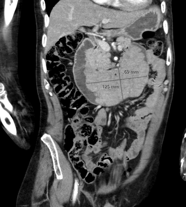
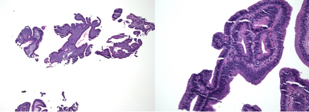
Methods: We report a rare case of a 58-year-old male with chronic hepatitis B infection who was incidentally found to have a giant TVA of the small intestine. The patient was admitted for healthcare-associated pneumonia and uncomplicated cystitis. A CT scan of the chest partially captured a mass in the small intestine, warranting an imaging of the abdomen that showed a large mass in the duodenum that extended to the jejunum and measured up to 12.5 x 6.9 cm, most compatible with malignancy, possibly lymphoma (image 1). The patient denied any related symptoms. An upper endoscopy with a tissue biopsy was done, revealing a large, frond-like, circumferential, and polypoid mass starting in the second part of the duodenum just distal to the ampulla and extending to the proximal jejunum, measuring approximately 25 cm in length. The lesion appeared, causing luminal narrowing without obstruction. Histology showed surface epithelium of the duodenum with dysplastic cytologic surface epithelial changes composed of nuclear enlargement, hyperchromasia, and pseudostratification typical for TVA without evidence of carcinoma (Image 2). Given the size and location of the lesion, surgical resection was planned. Discussion: This case represents the largest duodenal TVA reported in literature, highlighting the importance of early detection, which is crucial due to the malignancy risk associated with it.
Figure: Image 1: Abdomen CT scan-coronal section
Figure: Images 2: Histopathology of the duodenal biopsy
Disclosures: Farah Abuazzam indicated no relevant financial relationships. Bilind Ismail indicated no relevant financial relationships. Michael Chammany indicated no relevant financial relationships. Hussien Haidari indicated no relevant financial relationships. Hassan Khanani indicated no relevant financial relationships. Sathya Jaganmohan indicated no relevant financial relationships.
Farah Abuazzam, MD1, Bilind S. Ismail, MD1, Michael Chammany, DO2, Hussien Haidari, MD3, Hassan Khanani, MS4, Sathya Jaganmohan, MD5. P1993 - Giant Tubulovillous Adenoma of the Small Intestine as an Incidental Finding, ACG 2025 Annual Scientific Meeting Abstracts. Phoenix, AZ: American College of Gastroenterology.

 photo")