Lorraine Chong Tai, MD, Claudia De la Flor, DO, Yun-Yee Tsang, DO, Una Della-Pietra, DO Broward Health Medical Center, Fort Lauderdale, FL Introduction: A lymphangioma is a benign cystic tumor originating from the lymphatic system. They are a rare entity in adults, typically seen in the pediatric population. Given their rarity, non-specific presentation, and potential to mimic other intra-abdominal pathologies, abdominal lymphangiomas can be challenging to diagnose.
Case Description/
Methods: A 54-year-old male with a history of alcohol abuse, hypothyroidism, hyperlipidemia, and GERD presented with worsening left upper quadrant abdominal pain for 1 year. A prior Computed tomography (CT) of the abdomen/pelvis with Intravenous (IV) contrast showing a loculated fluid within the left upper quadrant of the abdomen measuring 14.3 x 9.2 x 12.2 cm and diffuse gastric thickening. His Carcinoembryonic antigen (CEA) was elevated at 7.2 ng/mL. Paracentesis showed a low Serum Ascites Albumin Gradient (SAAG) and 91.5% lymphocytes. An endoscopy revealed chronic gastritis with Helicobacter organisms present with extrinsic impression along the greater curve in the gastric fundus and proximal gastric body, confirming the mass was located outside of the stomach. He would later require an exploratory laparotomy revealing a 15cm cystic mass, which was excised. Pathology confirmed multiloculated cysts with dilated lymphatic channels consistent with a lymphangioma. The patient recovered well postoperatively and was subsequently discharged. Discussion: Abdominal lymphangiomas are rare, benign tumors believed to be congenital malformations of the lymphatic system. These tumors are often asymptomatic but may cause abdominal pain, distension, nausea, and sometimes a palpable mass. Imaging methods like ultrasound and CT are essential for initial evaluation, revealing cystic, multilocular lesions. However, the differential diagnosis is broad, including pancreatic pseudocysts, ascites, gastrointestinal stromal tumors (GIST), and lymphangiomas, which can be confirmed by fluid analysis showing lymphocytes. Definitive diagnosis relies on histopathological examination, revealing cysts lined by endothelial cells and containing lymphocytes. Surgical excision is the primary treatment, providing both diagnosis and therapeutic benefits. Conservative drainage offers temporary relief but carries risks of recurrence and infection. Our patient’s initial aspiration failed due to fluid reaccumulation, necessitating surgical resection. This case underscores the importance of considering abdominal lymphangiomas in the differential diagnosis of abdominal pain, especially with cystic masses.
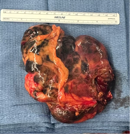
Figure: 15 cm wide abdominal lymphangioma after resection
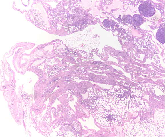
Figure: Histologic analysis showing multiloculated cysts with lymphoid aggregates
Disclosures: Lorraine Chong Tai indicated no relevant financial relationships. Claudia De la Flor indicated no relevant financial relationships. Yun-Yee Tsang: Enzolytics – Stocks that have been sold. Una Della-Pietra indicated no relevant financial relationships.
Lorraine Chong Tai, MD, Claudia De la Flor, DO, Yun-Yee Tsang, DO, Una Della-Pietra, DO. P1973 - A Huge Pain in the Guts: A Case of a Giant Abdominal Lymphangioma, ACG 2025 Annual Scientific Meeting Abstracts. Phoenix, AZ: American College of Gastroenterology.