Xavia Taylor, DO, Kelly Fuller, BS, Gregory Patterson, MD Archbold Medical Center, Thomasville, GA Introduction: Pyogenic granulomas (PGs), or lobular capillary hemangiomas, are benign vascular lesions commonly found on the skin and oral mucosa. Gastrointestinal involvement is exceedingly rare but clinically significant when present. In the small intestine, PGs can cause chronic bleeding, iron deficiency anemia, or serve as lead points for intussusception. Diagnosis is challenging due to their small size, vascularity, and frequent location beyond the reach of conventional endoscopy. Awareness of these atypical presentations is essential to ensure timely recognition and management.
Case Description/
Methods: A 78-year-old woman with hypertension, asthma, GERD, and chronic anemia presented with acute abdominal pain, nausea, and vomiting. CT revealed a target sign suggesting ileocecal intussusception and a 2 cm nodular mass near the cecum. Prior colonoscopy and EGD were negative. She underwent partial right colectomy and extended ileectomy. Pathology identified a large obstructing pyogenic granuloma. Her postoperative course included transfusion and a readmission for DVT and PE while anticoagulation was held. Hemoglobin stabilized with iron therapy, and she remains under hematology care. Discussion: While PGs are typically small and frequently missed on imaging or endoscopy, they may grow large enough to cause obstruction or act as a lead point for intussusception, as seen in this case. The etiology is unclear but may involve mucosal irritation or inflammation. This patient had recently completed H. pylori treatment, which has been associated with microbiome and immune changes in the ileocecal region. Early consideration of vascular lesions in cases of unexplained GI bleeding or obstruction may help avoid delayed diagnosis and improve outcomes.
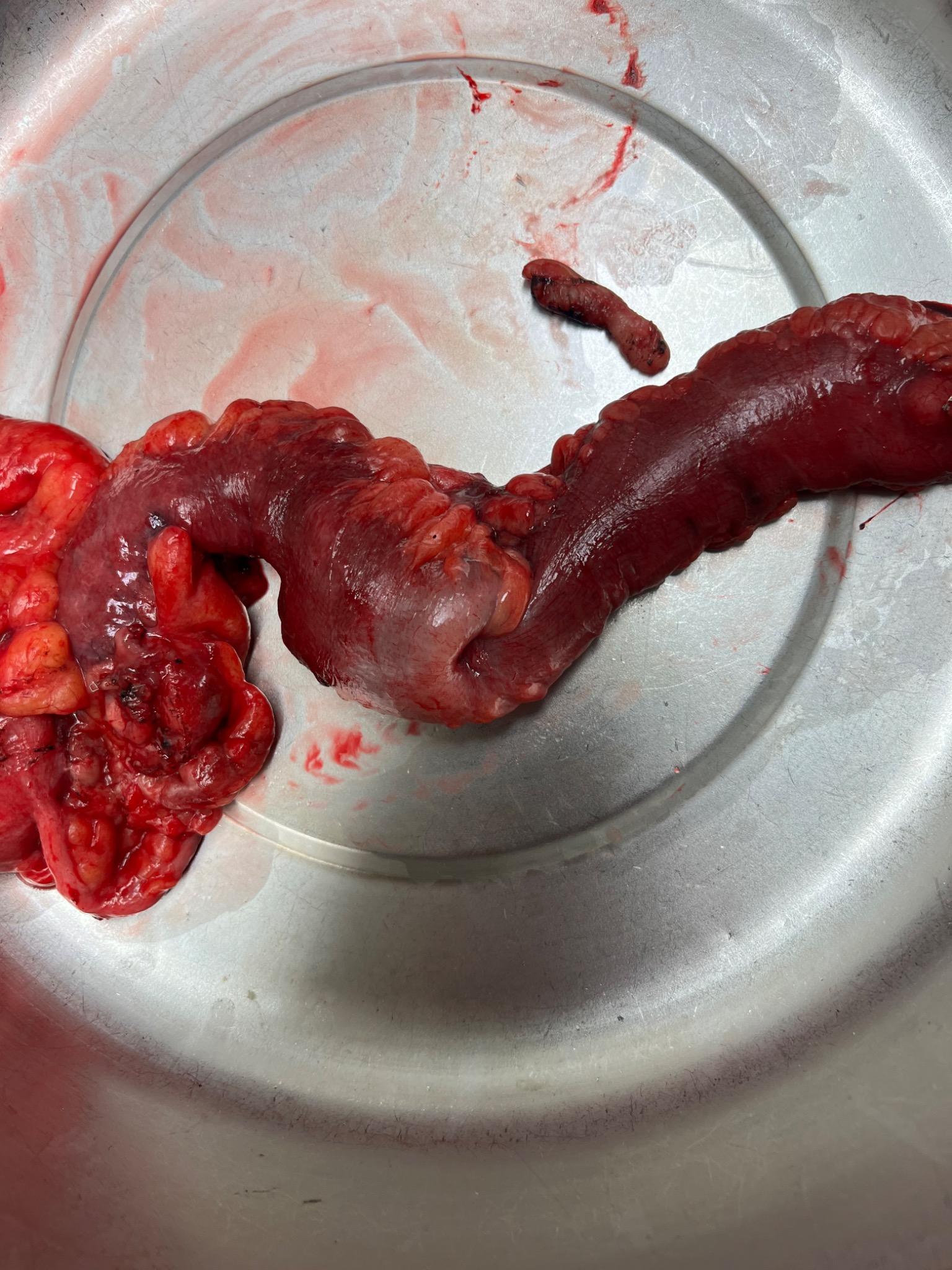
Figure: Gross pathology suggests intussusception, likely caused by the exophytic pyogenic granuloma. A well-circumscribed, lobulated mass with ulceration, hemorrhage, and necrosis is visible, consistent with pyogenic granuloma.
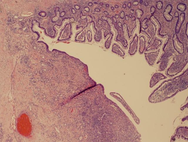
Figure: Histological section of small intestine showing a pyogenic granuloma (lobular capillary hemangioma), characterized by lobular clusters of capillaries within an inflamed stroma.
Disclosures: Xavia Taylor indicated no relevant financial relationships. Kelly Fuller indicated no relevant financial relationships. Gregory Patterson indicated no relevant financial relationships.
Xavia Taylor, DO, Kelly Fuller, BS, Gregory Patterson, MD. P1964 - Pyogenic Granuloma at the Ileocecal Junction: Unusual Culprit of Chronic Anemia and Intussusception, ACG 2025 Annual Scientific Meeting Abstracts. Phoenix, AZ: American College of Gastroenterology.

 photo")