University of North Carolina at Chapel Hill School of Medicine Chapel Hill, NC
Odai Mansour, BS1, Gianna Jones, BS1, Daniel Vanrooyen, DO2, Gabriel Winberry, MD3, Peter F. Farmer, MD4 1University of North Carolina at Chapel Hill School of Medicine, Chapel Hill, NC; 2Campbell University School of Osteopathic Medicine, Holly Springs, NC; 3Wakemed Pediatric Gastroenterology, Raleigh, NC; 4WakeMed Physician Practices, Raleigh, NC Introduction: The diagnosis of Collagenous Gastritis (CG) is made with esophagogastroduodenoscopy (EGD) biopsy revealing collagen deposition in the lamina propria of the gastric corpus. In pediatric patients, CG typically has a heterogeneous presentation with clinical signs and symptoms including moderate to severe anemia, weight loss, abdominal pain, diarrhea, and/or vomiting. CG is a rare disease and is likely underdiagnosed given the variable presentations. Existing literature characterizing CG and exploring the pathophysiology remains limited, contributing to a lack of definitive management strategies, uncertain prognosis and comorbid conditions, and impaired clinician awareness of diagnosis.
Case Description/
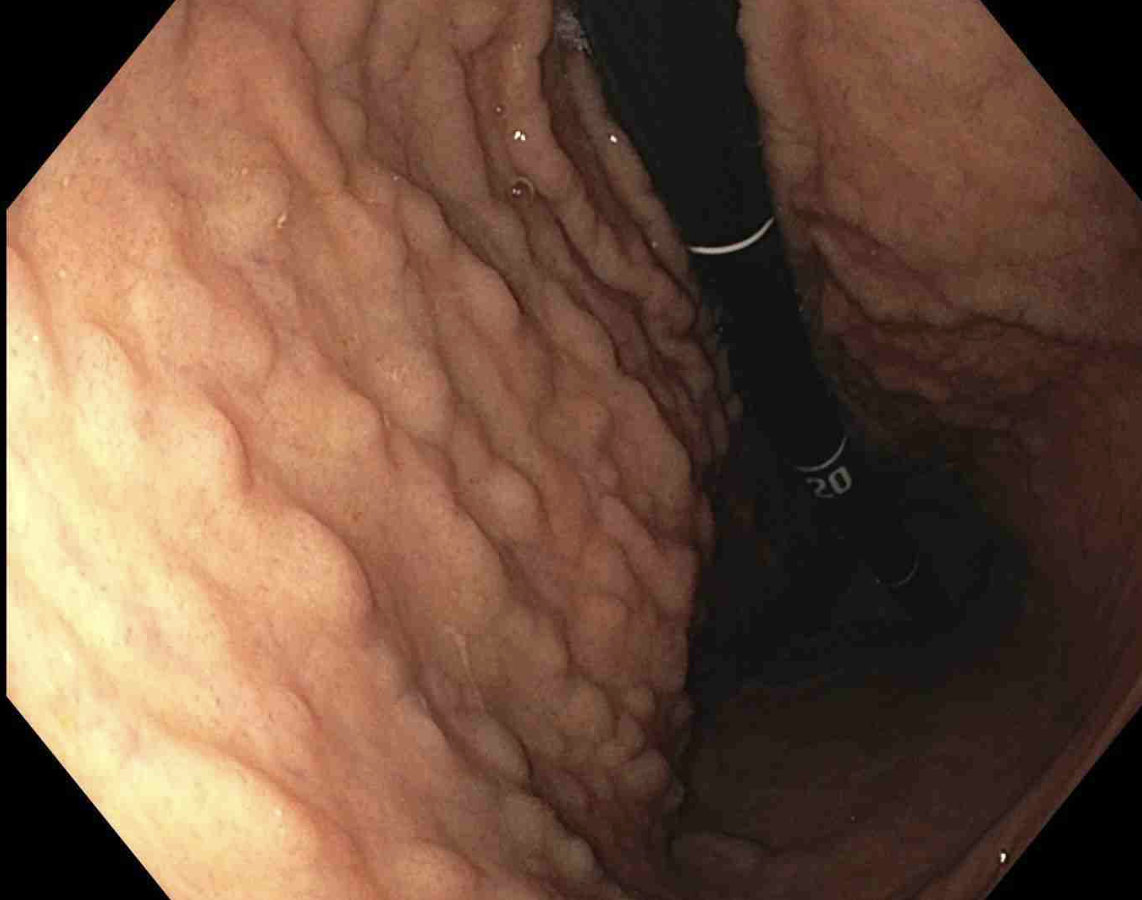
Methods: Patient 1 is a 13-year-old male who presented with recurrent headache, fatigue, poor weight gain with recent weight loss, cold intolerance, and pica. Although the patient denied abdominal pain, overt gastrointestinal bleeding, or changes to bowel patterns, EGD was performed (Figure 1) given a hemoglobin of 5.7 g/dL and elevated fecal calprotectin, and the biopsy confirmed CG (Figure 2).
Patient 2, an 8-year-old female, presented with intermittent non-bloody, non-bilious vomiting, non-bloody diarrhea, fatigue, and headaches. Workup revealed hemoglobin of 3.9 g/dL. Subsequent EGD with biopsy confirmed CG.
Patient 3, a 10-year-old male, initially presented with abdominal pain, nausea, vomiting, and poor weight gain and linear growth. Endoscopy with biopsy revealed moderate gastritis with increased eosinophils. Three years later, he was reevaluated for intense abdominal pain and was found to have mild microcytic anemia. EGD and biopsy confirmed CG.
Patient 4, a 17-year-old male, presented with refractory constipation, intermittent minor postprandial abdominal pain, and intermittent sensation of excessive fullness without particular food triggers. Lab work was notable only for historic iron deficiency anemia, previously diagnosed and managed with a multivitamin with iron. EGD with biopsy confirmed diagnosis of CG. Discussion: These four cases highlight the variable clinical signs and symptoms seen with collagenous gastritis. The most common sign across all cases appears to be moderate to severe anemia of unclear etiology. Research involving CG remains limited, and these four cases highlight the importance of considering CG when presented with moderate anemia of unknown origin in pediatric populations.
Figure: Figure 1. Endoscopic capture of the retroflex Gastric Body, demonstrating diffuse nodularity in Patient 1.
Figure: Figure 2. Histological biopsy of gastric mucosa under Masson’s Trichome Stain at 10x magnification, revealing subepithelial collagen banding and surface epithelial damage in Patient 1.
Disclosures: Odai Mansour indicated no relevant financial relationships. Gianna Jones indicated no relevant financial relationships. Daniel Vanrooyen indicated no relevant financial relationships. Gabriel Winberry indicated no relevant financial relationships. Peter Farmer indicated no relevant financial relationships.
Odai Mansour, BS1, Gianna Jones, BS1, Daniel Vanrooyen, DO2, Gabriel Winberry, MD3, Peter F. Farmer, MD4. P1905 - Pediatric Collagenous Gastritis: A Case Series, ACG 2025 Annual Scientific Meeting Abstracts. Phoenix, AZ: American College of Gastroenterology.