Washington University School of Medicine in St. Louis St. Louis, MO
Joseph Gung, MD1, Rachel O'Brien, ANPC2, Ige George, MD, MS1, Malak Itani, MD1, Vivian Ortiz, MD1 1Washington University School of Medicine in St. Louis, St. Louis, MO; 2Washington University School of Medcine in St. Louis, St. Louis, MO Introduction: Liver mass evaluation often requires a multidisciplinary team discussion for optimal management. When images are atypical, biopsy becomes essential to establish a diagnosis. Here, we describe a case of a 76-year-old female with diabetes mellitus type 2 and hypertension who was suspected to have malignancy in her liver until biopsy unfolded an atypical etiology.
Case Description/
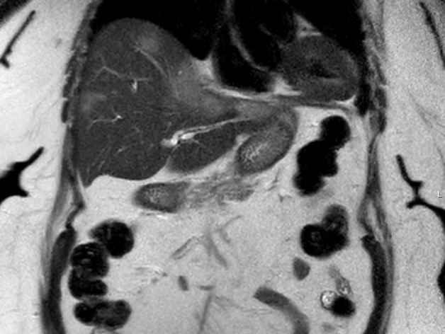
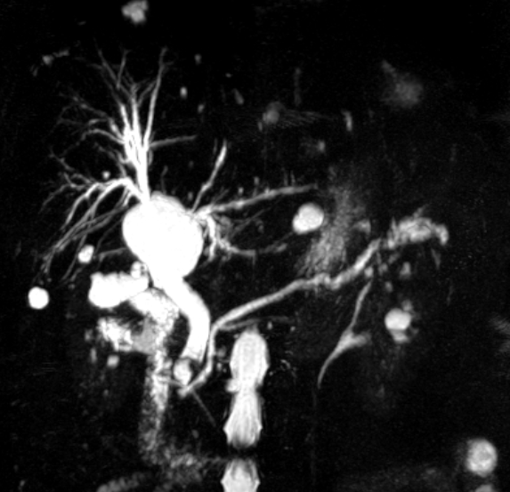
Methods: The patient presented with 2 weeks of persistent abdominal pain. Computer tomography of the abdomen showed multiple non-specific lesions throughout the liver, prominent common bile duct, and irregular configuration at the head of the pancreas. Follow up Magnetic Resonance Imaging (MRI) of the abdomen revealed redemonstrated scattered liver lesions with a dominant segment 2/4A mass measuring 6.9 x 3.2 cm suspicious for cholangiocarcinoma. Laboratory testing showed ALT 106, AST 65, ALP 436, Tot Bil 0.7, Alb 3.4, WBC 10.1, Hgb 12.8 and platelets 629. Endoscopic ultrasound identified a 10 x 4 mm mass versus stone in the common bile duct with no pancreas abnormalities, while periportal lymph node biopsy was negative for malignancy. For definitive diagnosis, ultrasound-guided liver biopsy was performed, which was complicated by acute hemorrhage immediately post-procedure requiring emergent coil embolization of segment 2/3 artery. Patient, however, subsequently developed septic shock from disseminated Mycobacterium fortuitum infection detected in the blood as well as liver abscesses growing multi-drug resistant Klebsiella pneumonia. Liver biopsy did not reveal acid-fast bacilli organisms or evidence of malignancy but she clinically improved on combined antibacterial and antimycobacterial therapy, with percutaneous drain placement. Drains were removed after two months, and antimicrobials continued for one year. Serial MRIs of her abdomen were obtained throughout her treatment showing resolution of liver masses and abscess at the end of her therapy. Discussion: Mycobacterium species infections mimicking liver malignancies have been rarely reported, and they are often associated with immunosuppressed host or underlying pulmonary disease, fish/aquatic-related exposures or direct inoculation. It is unclear how this patient acquired this infection. This case underscores the importance of biopsy in atypical hepatic lesions and the need for multidisciplinary management in complex hepatobiliary presentations.
Figure: MRI Abdomen series 36
Figure: MRI Abdomen series 3
Disclosures: Joseph Gung indicated no relevant financial relationships. Rachel O'Brien indicated no relevant financial relationships. Ige George indicated no relevant financial relationships. Malak Itani indicated no relevant financial relationships. Vivian Ortiz: Guerbet – Advisory Committee/Board Member.
Joseph Gung, MD1, Rachel O'Brien, ANPC2, Ige George, MD, MS1, Malak Itani, MD1, Vivian Ortiz, MD1. P1729 - Lucky Liver Mass, ACG 2025 Annual Scientific Meeting Abstracts. Phoenix, AZ: American College of Gastroenterology.