Rana Daas, MD, Michelle Rosario, MD University of South Florida, Tampa, FL Introduction: Lymphangiomas are benign mesenchymal tumors of the lymphatic system that occur on the skin and mucous membranes. They are caused by the dilation of lymphatic vessels resulting in cystic masses. Presentation may be asymptomatic to pain, pruritus, discharge, bleeding and iron deficiency anemia. Endoscopic findings include mucosal transparency and capillary elongation. Management typically involves monitoring, total surgical excision or abrasive therapies. They are rarely found in the anal and perianal region with few cases documented in the literature. In the perianal region, lymphangiomas can be mistaken for malignancy or other benign lesions highlighting the importance of recognition.
Case Description/
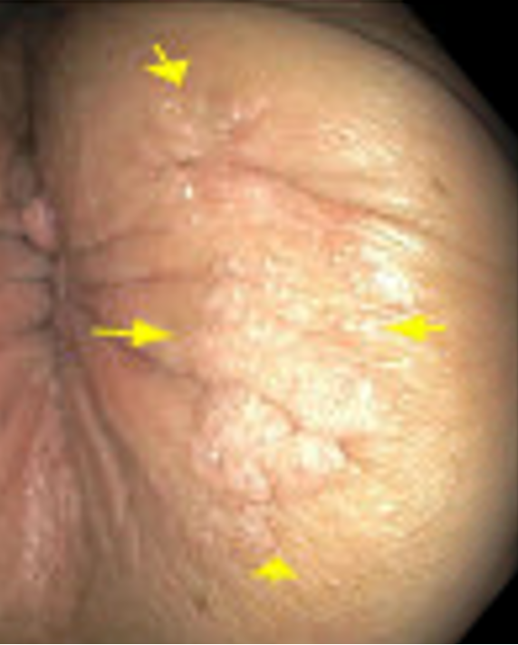
Methods: A 46-year-old female with a history of perianal fistulizing Crohn’s colitis requiring left hemicolectomy currently on vedolizumab and tofacitinib as well as multiple basal cell carcinomas presented for routine colonoscopy. She was found to have a 2 mm polyp in the transverse colon, no evidence of active Crohn’s, and a verrucous appearing lesion in the perianal region near her left buttock (Figures 1 & 2). The patient denied any perianal symptoms and was referred for surgical evaluation. Pathologic evaluation identified the lesion as a lymphangioma and no further intervention was recommended. Discussion: The detection of a perianal lesion in patients with fistulizing Crohn’s should prompt timely evaluation as chronic perianal fistulas can develop into malignancies including adenocarcinoma or squamous cell carcinoma. Additionally, verruciform lesion features raise concern for condyloma acuminatum due to human papilloma virus, verrucous carcinoma, and verruciform xanthoma. Although this patient has a history of perianal fistulizing disease and a lesion with verrucous features, her mass was determined to be a benign lymphangioma. Inflammatory bowel disease, including fistulizing Crohn’s, is a known trigger for acquired lymphangioma which can not only cause symptoms but also affect psychosexual function. Histopathologic evaluation is imperative to make a definitive diagnosis to guide management in these patients.
Figure: Figure 1: Perianal Lymphangioma.
Figure: Figure 2: Endoscopic remission of Crohn’s disease with simple endoscopic score of 0. Terminal ileum (A), cecum (B), transverse colon (C), and rectum (D).
Disclosures: Rana Daas indicated no relevant financial relationships. Michelle Rosario indicated no relevant financial relationships.
Rana Daas, MD, Michelle Rosario, MD. P1265 - A Rare Case of a Perianal Lymphangioma in Crohn’s Colitis, ACG 2025 Annual Scientific Meeting Abstracts. Phoenix, AZ: American College of Gastroenterology.

.jpg "Rana Daas, MD photo")