Huda Jaffar, MBBS, MD1, Umair Masood, MD2 1SUNY Upstate Medical University, Syracuse, NY; 2Mercy Hospital of Buffalo - Catholic Health System, Buffalo, NY Introduction: Colonoscopy is generally safe, however complications can occur rarely. A study showed patients >80 years having increased gastrointestinal adverse events and perforation risk1. We report a 64-year-old male presenting with umbilical and supraumbilical hernia incarceration after an uneventful routine colonoscopy.
Case Description/
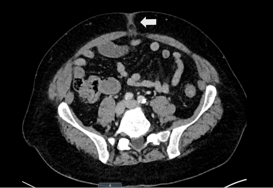
Methods: A 64-year-old man presented with a painful lump above his umbilicus a day after a routine colonoscopy. The patient had a history of diverticulitis evident on a recent CT scan and a bout of treated Clostridium difficile infection 3 months prior, and a colonoscopy was carried out to follow up on that. Procedure revealed mild colitis in the sigmoid colon and multiple polyps ranging from 3 to 25 mm that were removed via cold snare and cauterization. Diverticulosis was also noted, and a tattoo was placed in the sigmoid colon to mark the site of large piecemeal polypectomy. He subsequently developed pain above his umbilicus and arrived at the emergency department the next day. Clinical examination showed a small abdominal wall hernia, mildly tender on palpation. No laboratory derangements were noted apart from an elevated platelet count of 472 x 109/L (Reference range: 150-450 x 109/L). CT scan of the abdomen and pelvis revealed a small left abdominal wall hernia containing fluid with adjacent fat stranding suggesting incarceration. A laparoscopic central hernia repair was carried out. A 1-cm supraumbilical hernia with incarcerated preperitoneal fat was seen. Additionally, the surgery revealed a 1 cm umbilical hernia. A 9-cm symbotex mesh was used to cover both sites. Recovery was uneventful and he was discharged on the third day. Discussion: Hernia incarceration post-colonoscopy is rare. Prior reports involved female or cirrhotic patients2-4. Our case is the first of incarceration of a concurrent umbilical and supraumbilical hernia incareration post-colonoscopy in a male patient. Insufflation during the procedure needed to visualize and inspect the colonic mucosa may contribute to this. In our case, both air and CO2 were used due to limited distension. Generally, the risk is lower with CO2 compared to air because it is absorbed faster5. Patients at risk should undergo pre- and post-procedure abdominal examinations.
References:
Day LW et al., Gastrointest Endosc, 2011, p885–896
Beetham M et al., N Z Med J, 2009, p97–99
Al-shahrani A et al., Am J Gastroenterol, 2014, S465
Krawczyk M et al., Am J Med, 2015, pe13–e14
Hayman CV et al., World J Gastroenterol, 2021, p233–239
Figure: CT scan showing left abdominal wall hernia (white arrow) containing fluid with adjacent fat stranding, suggestive of incarceration
Disclosures: Huda Jaffar indicated no relevant financial relationships. Umair Masood indicated no relevant financial relationships.
Huda Jaffar, MBBS, MD1, Umair Masood, MD2. P0875 - An Unusual Case of Supraumbilical Hernia Incarceration After Colonoscopy, ACG 2025 Annual Scientific Meeting Abstracts. Phoenix, AZ: American College of Gastroenterology.