Orlando VA Medical Center and UCF/HCA Greater Orlando Kissimmee, FL
Adlin Lawrence, MD1, Paul Stolarov, BS2, Joane Titus, MD1, Nima Hosseinian, MD3 1Orlando VA Medical Center and UCF/HCA Greater Orlando, Kissimmee, FL; 2University of Central Florida College of Medicine, Orlando, FL; 3University of Central Florida, HCA Healthcare GME, Kissimmee, FL Introduction: Infectious esophagitis is a frequent complication in immunocompromised individuals, particularly those with AIDS. Candida albicans, cytomegalovirus (CMV), and herpes simplex virus (HSV) are the most common pathogens implicated in infectious esophagitis. Although endoscopic patterns can suggest a specific etiology, overlapping features may lead to diagnostic uncertainty. This case illustrates the importance of biopsy confirmation in guiding effective treatment.
Case Description/
Methods: A 25-year-old woman presented with progressive dyspnea, productive cough, and odynophagia. Examination revealed oral thrush and mucosal ulcers. Laboratory workup confirmed a new diagnosis of HIV/AIDS with a CD4 count of 24 cells/µL. Candida esophagitis was presumed, and fluconazole therapy was initiated. Despite treatment, odynophagia persisted. Upper endoscopy revealed erosive gastritis and deep, punched-out ulcers in the distal esophagus—features suggestive of CMV esophagitis. Elevated CMV IgG ( >8) led to initiation of ganciclovir. However, esophageal biopsies later identified HSV as the causative agent, and therapy was appropriately switched to acyclovir. Discussion: Esophagitis occurs in up to 30–50% of patients with AIDS, commonly due to Candida, CMV or HSV. While Candida esophagitis often responds to empirical antifungal therapy, CMV and HSV require pathogen-specific antivirals guided by accurate diagnosis. Endoscopic features—such as linear ulcers in CMV or vesicular lesions in HSV—can suggest an etiology but are not definitive. Studies show that biopsy offers over 90% sensitivity for distinguishing CMV from HSV, making it essential for diagnostic confirmation. In this case, HSV esophagitis presented with deep ulcers typically attributed to CMV, delaying the correct treatment. Similar delays are documented in the literature, where reliance on endoscopy alone led to suboptimal outcomes. Co-infections and overlapping features further complicate diagnosis in immunocompromised patients. Early biopsy with histopathologic and immunohistochemical analysis is crucial for accurate identification, enabling timely, targeted therapy and avoiding unnecessary broad-spectrum treatments, thereby improving clinical outcomes in this high-risk population.
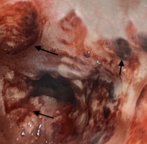
Figure: Figure 1: Endoscopy pictures showing deep, punched - out ulcers (black arrows) in the distal esophagus, suggestive of Cytomegalovirus
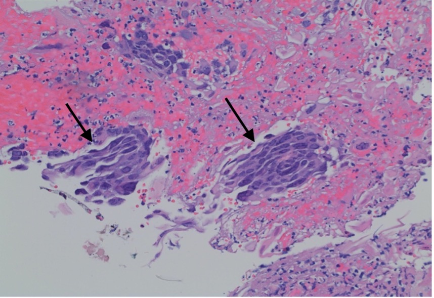
Figure: Figure 2: H&E stain of esophageal mucosa specimen at 20x magnification showing squamous mucosa with ulceration and viral cellular changes (black arrows), suggestive of HSV (Herpes simplex virus)
Disclosures: Adlin Lawrence indicated no relevant financial relationships. Paul Stolarov indicated no relevant financial relationships. Joane Titus indicated no relevant financial relationships. Nima Hosseinian indicated no relevant financial relationships.
Adlin Lawrence, MD1, Paul Stolarov, BS2, Joane Titus, MD1, Nima Hosseinian, MD3. P0761 - A Case of Misleading Findings: Atypical Presentation of HSV Esophagitis in AIDS, ACG 2025 Annual Scientific Meeting Abstracts. Phoenix, AZ: American College of Gastroenterology.