Crimean State Medical University named after Sal Georgivsky Simferopol, Ukraine Nawanshahr, Punjab, India
Neha Boski, MD, FRCR1, Parvinder Kaur, MD2, Mahendra Kumar, MBBS3, Nikhil Jain, MBBS4, Ujjwal Mishra, MBBS4, Rohan Raj, MBBS5 1Apollo Radiology International, Hyderabad, India, Hyderabad, Telangana, India; 2Crimean State Medical University named after Sal Georgivsky Simferopol, Ukraine, Nawanshahr, Punjab, India; 3Sardar Patel Medical College, Bikaner, India, Bikaner, Rajasthan, India; 4Uttar Pradesh University of Medical Sciences, Saifai, Etawah, Saifai, Uttar Pradesh, India; 5Memorial Hospital at Gulfport, Gulfport, MS Introduction: Dysphagia lusoria is classically described as difficulty swallowing due to esophageal compression by an aberrant right subclavian artery. Adult-onset cases are rare. We present a case of late-onset dysphagia in an elderly patient due to an aneurysmal aberrant left subclavian artery arising from a right-sided aortic arch.
Case Description/
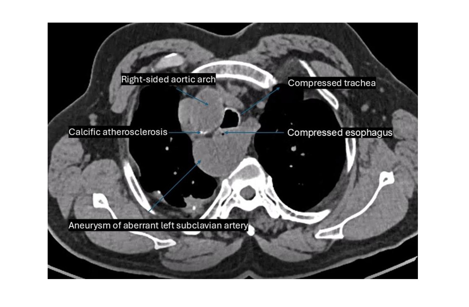
Methods: A 78-year-old male with a history of chronic obstructive pulmonary disease and hypertension presented with dysphagia to solids for one year and difficulty breathing. Chest X-ray revealed a high, right-sided aortic arch with tracheal compression. A subsequent barium swallow showed significant anterior displacement and focal luminal narrowing of the upper thoracic esophagus, suggesting a possible vascular ring. CT angiography of the chest was advised; however, due to impaired renal function, a non-contrast CT chest was performed. This revealed a right-sided aortic arch with an aberrant left subclavian artery and calcific atherosclerotic changes. An aneurysmal prominence of the proximal left subclavian artery was seen to exaggerate anterior displacement and compression over the esophagus and trachea. The cardiac apex was left-sided. Discussion: Delayed-onset dysphagia in the elderly due to a right-sided aortic arch with an aberrant left subclavian artery is rare. Tracheo-esophageal compression exacerbated by aneurysmal dilatation of the aberrant left subclavian artery is even rarer, warranting documentation of such cases. This case underscores the importance of considering vascular anomalies in the differential diagnosis of unexplained dysphagia in older adults. Barium swallow and cross-sectional imaging are essential tools for identifying extrinsic esophageal compression and defining the underlying vascular anatomy. Early recognition is critical for guiding appropriate management and preventing the progression of symptoms or related complications.
Figure: Axial non-contrast CT image of the upper thorax demonstrating a right-sided aortic arch with aneurysmal dilatation of the aberrant left subclavian artery causing significant compression of the esophagus and trachea.
Disclosures: Neha Boski indicated no relevant financial relationships. Parvinder Kaur indicated no relevant financial relationships. Mahendra Kumar indicated no relevant financial relationships. Nikhil Jain indicated no relevant financial relationships. Ujjwal Mishra indicated no relevant financial relationships. Rohan Raj indicated no relevant financial relationships.
Neha Boski, MD, FRCR1, Parvinder Kaur, MD2, Mahendra Kumar, MBBS3, Nikhil Jain, MBBS4, Ujjwal Mishra, MBBS4, Rohan Raj, MBBS5. P0751 - Adult-Onset Dysphagia Lusoria in a Right-Sided Aortic Arch With Aneurysm of the Aberrant Left Subclavian Artery, ACG 2025 Annual Scientific Meeting Abstracts. Phoenix, AZ: American College of Gastroenterology.

 photo")