Rishika Trivedi, MD1, Prince Shah-Riar, MD2, Praveen Vijhani, MD1, Asif Zamir, MD, FACG3, Murthy Badiga, MD, FACG4 1DHR Health, McAllen, TX; 2DHR Health, Edinburg, Tx, McAllen, TX; 3DHR Health Gastroenterology, Edinburg, TX; 4DHR Health Gastroenterology, McAllen, TX Introduction: Mallory-Weiss tears are longitudinal mucosal lacerations typically located at the gastroesophageal junction, commonly measuring between 0.75 and 1.5 inches (approximately 2–4 cm) in length . These lesions often result from forceful retching or vomiting and are generally self-limiting, with most cases resolving spontaneously or requiring minimal intervention . However, extensive esophageal mucosal tears exceeding 10 cm are exceptionally rare and not well documented in medical literature. We present a case of a massive 10 cm mid-esophageal mucosal tear managed successfully with endoscopic clipping and intensive supportive care.
Case Description/
Methods: A 62-year-old woman with hypertension, diabetes mellitus, coronary artery disease, and gastroparesis presented with sudden-onset, intractable nausea and vomiting followed by more than ten episodes of bright red hematemesis. She reported severe chest and epigastric pain following retching. Initial hemoglobin was 12.6 g/dL. Emergent esophagogastroduodenoscopy (EGD) revealed a large, long, and deep mucosal tear in the mid-esophagus measuring approximately 10 cm with active oozing and bleeding. The tear was closed using ten hemostatic clips (Boston Scientific) with successful hemostasis and no immediate complications (Figure 1). Clotted blood was found in the cardia, fundus, and body of the stomach; the duodenum was normal.
Post-procedure, the patient was admitted to the intensive care unit, sedated, and intubated for airway protection. CT chest and abdomen showed no pneumomediastinum or perforation. She remained NPO for one week per gastroenterology recommendation. A PICC line was placed and total parenteral nutrition (TPN) initiated. She remained hemodynamically stable with no signs of rebleeding, was successfully extubated the next day, and was later downgraded to the medical floor in improving condition. Discussion: This case highlights a rare, large esophageal mucosal tear treated entirely via endoscopic means. While most Mallory-Weiss tears are < 3 cm and located at the gastroesophageal junction, this case involved a 10 cm tear in the mid-esophagus. Timely EGD and placement of ten hemostatic clips allowed for complete closure and control of hemorrhage. ICU-level supportive care, including temporary mechanical ventilation, NPO status, and nutritional support with TPN, contributed to a favorable outcome. This case illustrates that even extensive esophageal mucosal injuries can be successfully managed without surgical intervention.
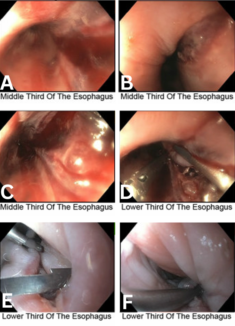
Figure: Figure 1. Endoscopic images showing a 10 cm mid-esophageal mucosal tear (A–C) with active oozing and clotted blood. Hemostasis was achieved with placement of ten hemostatic clips (D–F), with complete closure of the defect.
Disclosures: Rishika Trivedi indicated no relevant financial relationships. Prince Shah-Riar indicated no relevant financial relationships. Praveen Vijhani indicated no relevant financial relationships. Asif Zamir indicated no relevant financial relationships. Murthy Badiga indicated no relevant financial relationships.
Rishika Trivedi, MD1, Prince Shah-Riar, MD2, Praveen Vijhani, MD1, Asif Zamir, MD, FACG3, Murthy Badiga, MD, FACG4. P0740 - Massive Mid-Esophageal Mucosal Tear (10 cm) Managed Endoscopically With Hemoclips: A Case Report, ACG 2025 Annual Scientific Meeting Abstracts. Phoenix, AZ: American College of Gastroenterology.