Texas Tech University Health Science Center El Paso El Paso, TX
Mostafa Eysha, MD1, Mohanad Elchouemi, BS2, Omar Alkasabrah, MD3, Tushar Talaparthy, BS4, Alejandro Robles, MD5, Sherif E. Elhanafi, MD6 1Texas Tech University Health Science Center El Paso, El Paso, TX; 2Paul L. Foster School of Medicine, Texas Tech University Health Sciences Center, El Paso, TX; 3Landmark Medical Center, Woonsocket, RI; 4Tilman J. Fertitta Family College of Medicine, Houston, TX; 5Department of Gastroenterology, Paul L. Foster School of Medicine, Texas Tech University Health Sciences Center El Paso, El Paso , TX, El Paso, TX; 6Texas Tech University Health Sciences Center, El Paso, TX Introduction: Esophageal leiomyosarcoma is a rare malignant tumor accounting for 0.1 to 0.5% of diagnosed esophageal malignancies. It commonly occurs in the 5th to 7th decades of life. Patients typically present with dysphagia, retrosternal chest pain, and weight loss. We present a case of a 70-year-old male with esophageal leiomyosarcoma with unusual presentation of early metastases and symptomatic hypercalcemia.
Case Description/
Methods: A 70-year-old male presented with a 3-week history of constipation associated with decreased oral intake. The patient experienced fever, abdominal pain, a 16-pound weight loss, and stabbing chest pain exacerbated by movement. Patient denied dysphagia to solid or liquids. Laboratory workup revealed hypercalcemia (Ca2+ 12.6), low PTH 4.4. C-reactive protein, UPEP, SPEP, PTHrP, and 25 hydroxy-vitamin D were all within normal limits. Computed tomography of the chest and abdomen revealed a large mass in the distal esophagus, peri-celiac lymphadenopathy likely metastatic, and a hypodense 1.7 cm liver lesion, which was highly concerning for malignancy with metastases. Barium esophagogram suspected a large tumor at the gastroesophageal junction. Esophagogastroduodenoscopy was performed and showed a large fungating mass in the lower third of the esophagus protruding into the stomach (Siewart classification type 1). Biopsies were obtained and histology showed high grade leiomyosarcoma. Tumor cells were positive for smooth muscle actin and vimentin immunostains; CD138 immunostaining demonstrates nonspecific aberrant staining in tumor cells; tumor cells negative for desmin, myogenin, p40, CAM5.2, pan-keratin, CD117, CD43, CD3, CD1a, CD21, CD34, CD79a, TdT, CD20, S100, MUM1 and LCA immunostains. Ki-67 immunostaining demonstrates a high proliferative index (approximately 85% of tumor cells). The patient was treated for hypercalcemia with zoledronic acid and intravenous hydration with complete resolution biochemically and clinically. Oncology was consulted and recommended palliative chemotherapy due to the metastatic presentation.
Discussion: This case highlights that esophageal leiomyosarcoma can present with early metastasis even in the absence of dysphagia. The most frequent sites of metastasis include; bone, liver, and lungs. Early diagnosis and treatment is crucial as surgical resection in esophageal limited disease is associated with favorable surgical and oncologic outcomes.
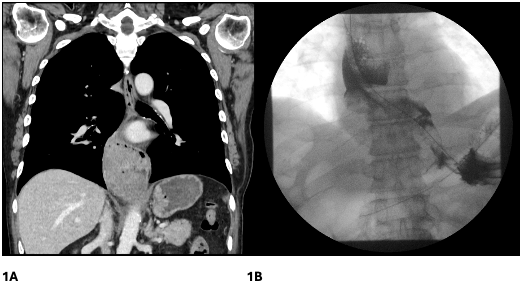
Figure: Figure 1A. Computed tomography of the chest and abdomen revealed a large mass in the distal esophagus, peri-celiac lymphadenopathy likely metastatic, and a hypodense 1.7 cm liver lesion.
Figure 1B. Barium esophagogram showing a large tumor at the gastroesophageal junction.
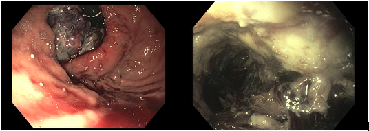
Figure: Figure 2. Esophagogastroduodenoscopy was performed and showed a large fungating mass in the lower third of the esophagus protruding into the stomach (Siewart classification type 1)
Disclosures: Mostafa Eysha indicated no relevant financial relationships. Mohanad Elchouemi indicated no relevant financial relationships. Omar Alkasabrah indicated no relevant financial relationships. Tushar Talaparthy indicated no relevant financial relationships. Alejandro Robles indicated no relevant financial relationships. Sherif Elhanafi indicated no relevant financial relationships.
Mostafa Eysha, MD1, Mohanad Elchouemi, BS2, Omar Alkasabrah, MD3, Tushar Talaparthy, BS4, Alejandro Robles, MD5, Sherif E. Elhanafi, MD6. P0731 - A Rare Case of Metastatic Esophageal Leiomyosarcoma Presenting as Symptomatic Hypercalcemia, ACG 2025 Annual Scientific Meeting Abstracts. Phoenix, AZ: American College of Gastroenterology.