Centro de Gastroenterologia Avanzada Santo Domingo, Distrito Nacional, Dominican Republic
Priscilla M. Guerrero-Camarena, MD1, Diego Abreu Delgado, MD1, Isabella Mella, MD1, Nicole Burgos, MD1, Pablo Socias-Pappaterra, MD2, Maria Isabel Cano, MD1, Albiriza Monegro, MD1, Cynthia Contreras, MD3, Elizabeth Tavarez, MD4, Fernando Contreras, MD, FACG1, Carlos J. Esteva, 5 1Centro de Gastroenterologia Avanzada, Santo Domingo, Distrito Nacional, Dominican Republic; 2Department of Knowledge Management and Epidemiology, Centros de Diagnóstico y Medicina Avanzada y de Conferencias Médicas y Telemedicina (CEDIMAT), Santo Domingo, Dominican Republic., Santo Domingo, Distrito Nacional, Dominican Republic; 3University of Kansas Medical Center, Kansas City, KS, Kansas City, KS; 4Centro de Diagnostico Medicina Avanzada y Telemedicina, Santo Domingo, Distrito Nacional, Dominican Republic; 5ESTEVA Patologia, Santo Domingo, Distrito Nacional, Dominican Republic Introduction: Sebaceous glands are microscopic, sac-like structures primarily found in the dermis, often associated with hair follicles composing pilosebaceous units that produce and store sebum. While commonly located in the skin, ectopic sebaceous glands have been found in the mouth, eyes, palms, soles, and even tongue.
Sebaceous Glands Metaplasia (SGM) is a unique condition where sebaceous glands are found on the surface of the esophageal mucosa, often discovered as an incidental finding during endoscopic evaluations for unassociated conditions. Studies suggest that SGM has an incidence of around 0.005% and can frequently be misdiagnosed for candidiasis, xanthomas, or squamous papillomas. We present a patient with ectopic sebaceous glands in the esophagus identified during routine endoscopy.
Case Description/
Methods: 40-year-old woman with no significant medical history presented with one month of reflux, which improved with over-the-counter proton pump inhibitors but worsened at night, accompanied by odynophagia and dyspnea. She reported a 10-year history of tobacco use and consumed one glass of wine daily until symptom onset. Physical examination and routine laboratory tests were unremarkable. Upper endoscopy revealed multiple small, lobulated pale plaques with a central punctate protrusion. These lesions were observed extending from 2 cm above the gastroesophageal junction to the mid-esophagus, appearing in patches and occasionally in linear patterns. Biopsies results revealed ectopic sebaceous glands.
Despite acid reflux and odynophagia being the patient's primary complaints, no evidence of esophagitis was found. The only notable findings were the ectopic sebaceous glands. Discussion: Ectopic sebaceous glands in the esophagus are rare and incidental findings, as they are generally asymptomatic. While our patient presented with non-specific symptoms, including odynophagia and reflux, there is no established association between of these and the presence of ectopic sebaceous glands. Furthermore, there were no notable endoscopic changes that could account for her presenting symptoms. The exact pathogenesis remains unclear, but proposed mechanisms include metaplastic changes or congenital anomalies. Recognizing this condition is important, as yellow esophageal plaques can mimic other entities, such as carcinoid tumors, granular cell tumors, or xanthomas. Existing literature indicates that ectopic sebaceous glands are generally benign, with a favorable prognosis and no need for treatment beyond diagnosis.
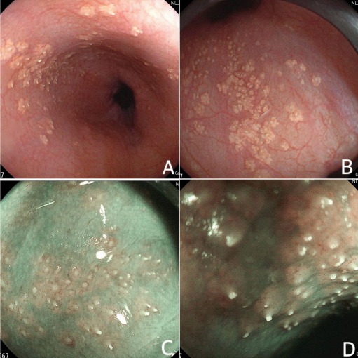
Figure: Figure 1. A,B,C,D. Multiple small, lobulated, pale plaques with central punctate protrusions were observed in the esophagus during upper endoscopy.
Figure: Figure 2. Esophageal biopsy revealing esophageal squamous mucosa with the presence of ectopic sebaceous glands.
Disclosures: Priscilla Guerrero-Camarena indicated no relevant financial relationships. Diego Abreu Delgado indicated no relevant financial relationships. Isabella Mella indicated no relevant financial relationships. Nicole Burgos indicated no relevant financial relationships. Pablo Socias-Pappaterra indicated no relevant financial relationships. Maria Isabel Cano indicated no relevant financial relationships. Albiriza Monegro indicated no relevant financial relationships. Cynthia Contreras indicated no relevant financial relationships. Elizabeth Tavarez indicated no relevant financial relationships. Fernando Contreras indicated no relevant financial relationships. Carlos Esteva indicated no relevant financial relationships.
Priscilla M. Guerrero-Camarena, MD1, Diego Abreu Delgado, MD1, Isabella Mella, MD1, Nicole Burgos, MD1, Pablo Socias-Pappaterra, MD2, Maria Isabel Cano, MD1, Albiriza Monegro, MD1, Cynthia Contreras, MD3, Elizabeth Tavarez, MD4, Fernando Contreras, MD, FACG1, Carlos J. Esteva, 5. P0730 - Esophageal Acne: An Incidental Finding in Oral Endoscopy, ACG 2025 Annual Scientific Meeting Abstracts. Phoenix, AZ: American College of Gastroenterology.