Lorraine Chong Tai, MD1, Haille Joseph, MBBS2 1Broward Health Medical Center, Fort Lauderdale, FL; 2Scarborough General Hospital, Signal Hill, Tobago, Trinidad and Tobago Introduction: Cervical osteophytes are bony outgrowths or bone spurs of the cervical vertebrae. They are a common degenerative finding, particularly in the elderly population. Anterior cervical osteophytes are present in approximately 20-30% of the elderly population. Dysphagia resulting from cervical osteophytes is a rare but critical diagnosis to consider, as delayed identification can lead to complications like malnutrition, aspiration, or obstruction.
Case Description/
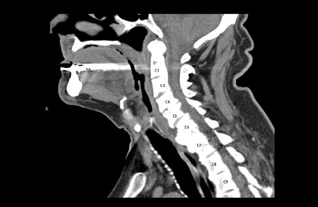
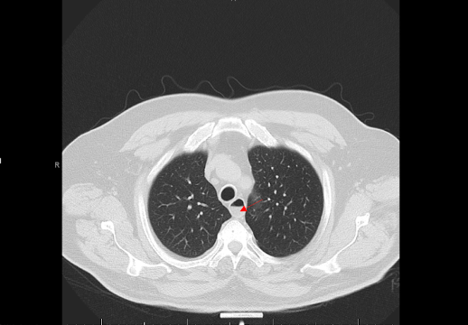
Methods: We present the case of a 67-year-old male who presented to the emergency department for food impaction after he was eating a hotdog. He had associated drooling with one episode of vomiting and dysphagia to liquids. He endorsed having a similar episode several years prior that did not require any endoscopic intervention. Initial labs and vitals were unremarkable. A Computed tomography (CT) scan of the neck showed a prominent anterior osteophyte at C6-C7 vertebrae (Figure 1) causing some mass effect of the esophagus and a CT scan of the chest (Figure 2) showed a soft tissue density in the distal esophagus consistent with food impaction. He was admitted and started on Intravenous(IV) Fluids, IV Protonix, kept nothing by mouth(NPO) and Gastroenterology was consulted. Overnight his food bolus passed, and he was able to drink water without difficulty. No endoscopic intervention was required, and he was subsequently discharged with recommendations to follow up for an osteophytectomy. Discussion: Cervical osteophytes are often seen in those greater than 60 years of age and thought to develop due to the constant strain or heavy lifting resulting in hypertrophy of the cervical bones forming bone spurs. The mechanism of dysphagia is thought to involve mechanical obstruction, inflammation of surrounding soft tissues leading to fibrosis, impaired function of the esophageal muscles due to direct compression or cricopharyngeal spasm triggered by pressure on the esophagus. It has also been suggested that dysphagia in patients with osteophytes can result from the inability of the epiglottis to properly tilt over the laryngeal inlet due to osteophytes at the C3 and C4 levels. Management can range from conservative measures, such as dietary modifications and anti-inflammatory medications, to surgical intervention in severe cases. Radiological imaging is essential for diagnosis, and osteophytectomy is an effective surgical option. Early detection and management can greatly improve patient outcomes.
Figure: Figure 1: Contrast-enhanced CT scan of the neck showed a prominent anterior osteophyte at C6-C7 vertebrae causing some mass effect of the esophagus
Figure: Figure 2: CT scan of the chest showing a red arrow indication a soft tissue density in the distal esophagus consistent with food impaction
Disclosures: Lorraine Chong Tai indicated no relevant financial relationships. Haille Joseph indicated no relevant financial relationships.
Lorraine Chong Tai, MD1, Haille Joseph, MBBS2. P0716 - Bone Appétit: Anterior Cervical Osteophyte Leading to Food Impaction, ACG 2025 Annual Scientific Meeting Abstracts. Phoenix, AZ: American College of Gastroenterology.