Angela Xue, MD1, Firrah Saeed, MD1, Jennifer Katz, MD, FACG2 1NYU Langone Health, New York, NY; 2NYU Grossman School of Medicine, New York, NY Introduction: Percutaneous endoscopic gastrostomy (PEG) is the preferred modality for enteral nutrition in patients with limited oral intake.¹ Ultrasound-assisted PEG placement should be considered in patients with challenging anatomy to ensure safe tube placement.
Case Description/
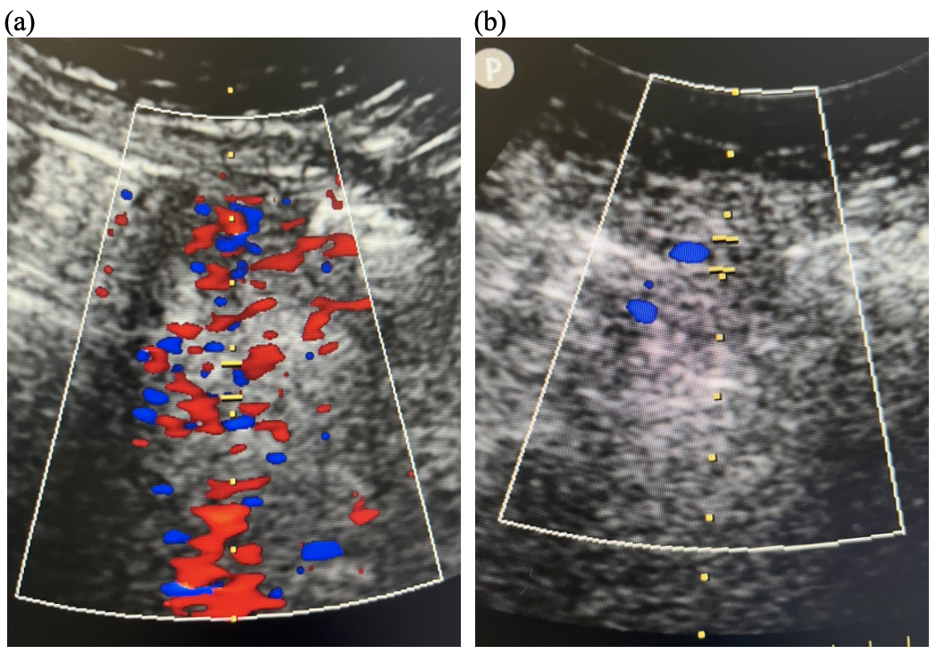
Methods: Mr. R, a 59-year-old male with hypertension, hyperlipidemia, and type 2 diabetes mellitus, presented with acute left-sided weakness and subsequent unresponsiveness. Imaging revealed a right pontine hemorrhage, necessitating intubation and nasogastric tube placement. Given ongoing enteral nutrition needs, Gastroenterology was consulted for PEG placement. A CT abdomen/pelvis without IV contrast suggested mesenteric vessels anterior to the stomach, raising concern for vascular injury risk. Therefore, ultrasound-assisted PEG placement was pursued. After appropriate gastric insufflation, a site in the stomach body demonstrated excellent transillumination and external indentation. Ultrasound identified pulsatile blood flow along portions of the abdominal wall. A safe zone without vascular structures was marked. A 20 Fr Boston Scientific EndoVive Safety gastrostomy tube was successfully placed using the pull method. The patient tolerated tube feeds post-procedure without complications. Discussion: Standard PEG placement typically does not require ultrasound guidance in average-risk patients when proper techniques are applied. However, in high-risk anatomical scenarios, ultrasound can mitigate procedural bleeding risks. This case highlights the utility of ultrasound in identifying aberrant or adjacent vasculature, preventing potential complications. Prior reports support ultrasound-assisted PEG placement in patients with abdominal wall varices, hepatomegaly, or coagulopathy.²˒³ Gastroenterologists should consider ultrasound guidance in patients with altered anatomy or increased vascular proximity to enhance procedural safety.
1. Vudayagiri L, et al. Percutaneous Endoscopic Gastrostomy Tube, 2025, StatPearls Publishing. 2. Sanchez V. Utility of Ultrasound-Assisted Percutaneous Gastrostomy in Anticoagulated Patients with Hepatomegaly, 2022, ACS. 3. Höroldt BS, et al. Ultrasound guidance in PEG placement: An adjuvant technique in patients with abdominal wall varices? Dig Liver Dis. 2005;37(9):709–712.
Figure: Figure 1. Doppler Ultrasound of Abdominal Cavity. (a). Site with extensive vasculature. (b). Procedural site with minimal vasculature.
Disclosures: Angela Xue indicated no relevant financial relationships. Firrah Saeed indicated no relevant financial relationships. Jennifer Katz indicated no relevant financial relationships.
Angela Xue, MD1, Firrah Saeed, MD1, Jennifer Katz, MD, FACG2. P0573 - Avoiding a Vascular Misadventure: The Case for Ultrasound-Assisted PEG, ACG 2025 Annual Scientific Meeting Abstracts. Phoenix, AZ: American College of Gastroenterology.

 photo")