
Sunday Poster Session
Category: Colon


Casey M. DeBeltz, DO
CHI Health Creighton University Medical Center
Omaha, NE
Bowel intussusception cases are more commonly diagnosed in pediatric patients, and in these cases, are typically related to benign etiology. Intussusception is rare in adults. When diagnosed in the large intestine, they are typically related to tumors, polyps, or inflammatory lesions. We present an interesting case of colonic intussusception in an adult female not related to malignancy or inflammatory bowel disease.
Case Description/
Methods:
A 54-year-old female with chronic constipation and recurrent uncomplicated diverticulitis presented with worsening nausea and abdominal pain. Initial CT scan was concerning for a sigmoid “stricture” with proximal colonic distention, similar to imaging from several years prior. Repeat CT with rectal gastrografin revealed a characteristic stenotic “apple core lesion” concerning for malignancy. She underwent an inpatient colonoscopy which demonstrated congested mucosa in the mid-sigmoid colon suggestive of sigmoid intussusception without fixed stenosis or stricture. Mucosa from this area was biopsied and was negative for inflammatory changes or malignancy. Of note, colonoscopy with biopsy of the area of interest four years prior had similar findings. Because of recurrent presentations for diverticulitis and obstructive symptoms, the patient underwent a sigmoid colectomy with diverting loop ileostomy. She was found to have intrabdominal adhesions and several fibrotic nodules and scarring on the outer surface of the colon leading to a tight stricture. Surgical pathology showed multiple diverticula but was negative for malignancy.
Discussion:
This case illustrates the rare occurrence of stenosis due to recurrent diverticulitis as a lead point for intussusception. More common causes of intussusception in adults such as tumors, polyps, adhesions, or inflammatory lesions were ruled out in our patient based on endoscopic imaging, endoscopic biopsies, intraoperative findings, and final surgical pathology. 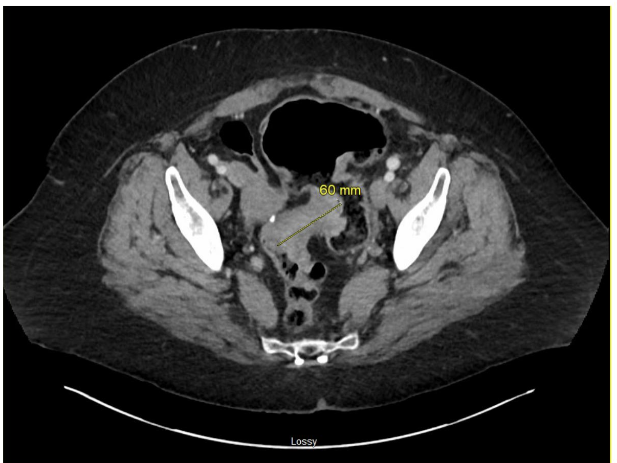
Figure: CT scan with rectal gastrograffin showing “apple core lesion” concerning for malignancy in the sigmoid colon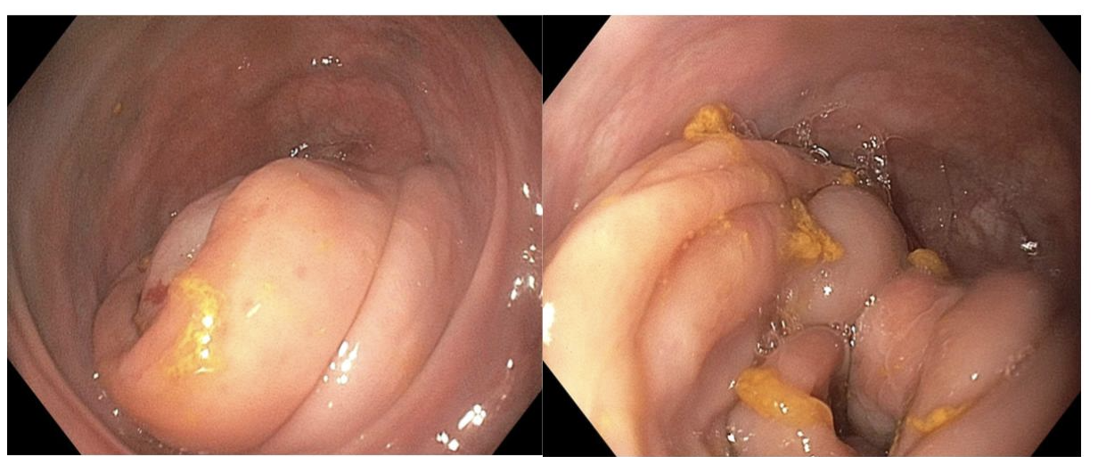
Figure: Colonoscopy images reveal congested mucosa in the sigmoid colon
Disclosures:
Casey DeBeltz indicated no relevant financial relationships.
Clive Miranda indicated no relevant financial relationships.
Fnu Monika indicated no relevant financial relationships.
Aditya Avula indicated no relevant financial relationships.
Marcellus Singh indicated no relevant financial relationships.
Robert Kizer indicated no relevant financial relationships.
Casey M. DeBeltz, DO, Clive J. Miranda, DO, MSc, Fnu Monika, MBBS, Aditya Avula, DO, Marcellus Singh, MD, Robert Kizer, MD. P0348 - Chronic Diverticula as a Lead Point in Adult Sigmoid Colon Intussusception, ACG 2025 Annual Scientific Meeting Abstracts. Phoenix, AZ: American College of Gastroenterology.