Sunday Poster Session
Category: Colon
Raehun Lee, MD
Creighton University School of Medicine
Phoenix, AZ
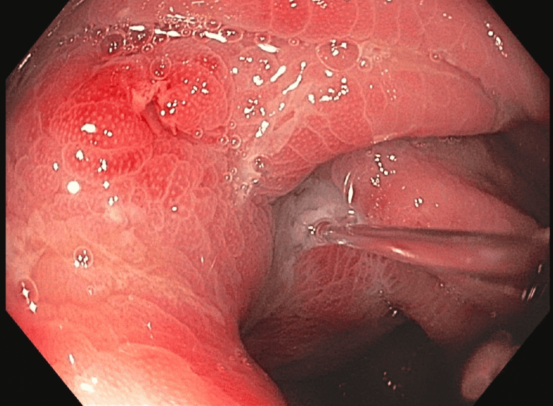
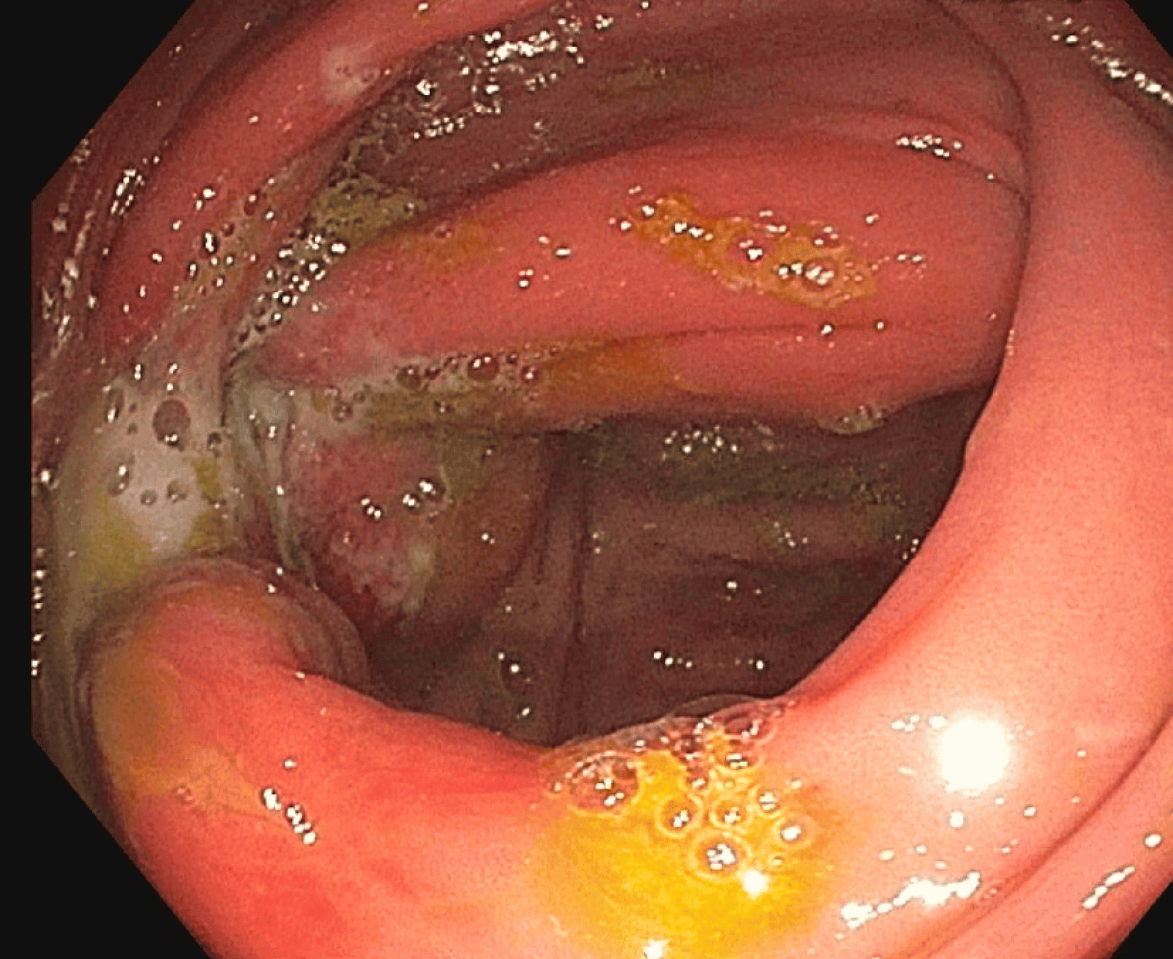
Cecal diverticulitis is a rare cause of right lower quadrant pain in Western populations and often mimics appendicitis. CT imaging is essential for accurate diagnosis and can help avoid unnecessary surgery. This case demonstrates how imaging guided the diagnosis and urgent colonoscopic evaluation of a suspected complicated cecal diverticulitis.
A 48-year-old male with no significant past medical history presented with acute right lower quadrant abdominal pain and stable vital signs. Laboratory workup showed leukocytosis (WBC 12.3), elevated inflammatory markers (ESR 45, CRP 136), and fecal calprotectin of 604 µg/g. Initial CT abdomen/pelvis with IV contrast revealed a 4.5 cm debris-filled outpouching from the anterior cecum, with wall thickening and pericolonic fat stranding. Differential at the time included acute right-sided diverticulitis versus contained perforation of a cecal mass. A follow-up CT with oral contrast confirmed similar findings, prompting urgent colonoscopy due to the concern for an evolving perforation and to obtain tissue diagnosis.
Right-sided diverticulitis is uncommon in Western populations and often mimics acute appendicitis, frequently leading to unnecessary surgical intervention. In this case, CT imaging was instrumental in identifying a large inflammatory cecal outpouching with normal appendix, shifting the diagnostic focus away from appendicitis. This radiologic clarity enabled a conservative, non-surgical approach with antibiotics and close follow-up. Urgent colonoscopy, guided by imaging, confirmed the source of inflammation and excluded malignancy. This case highlights how early and accurate imaging can prevent unwarranted surgery by distinguishing diverticulitis from surgical mimics.