Corey Garcia, 1, Amanda Eukovich, DO2, Ivana Rubenstein, DO2, Lorraine Chong Tai, MD2, Edwin Makarevich, DO2, Satya Singh, MD2 1Nova Southeastern University, Royal Palm Beach, FL; 2Broward Health Medical Center, Fort Lauderdale, FL Introduction: A giant gallbladder (GGB) is a rare condition defined by: length > 14cm, a volume > 1.5L, or a weight > 1.5kg. It is usually associated with obstructive stones, chronic cholecystitis, neoplasms, adhesions, bacterial infections of the gallbladder, and functional gallbladder disorders. Herein we describe a case of a young patient with a 18.5x6.27x4.39cm gallbladder in the setting of a type IVa choledochal cyst (CC4A) and primary sclerosing cholangitis (PSC).
Case Description/
Methods: A 19 year-old male with a past medical history of primary sclerosing cholangitis (PSC) presented to the ED for evaluation of 3 weeks of jaundice, scleral icterus and darkening of his urine. CT imaging showed severe intrahepatic biliary ductal dilatation, severe common bile duct (CBD) dilatation (2.2cm), and a severely distended gallbladder with debris/stones in the distal CBD. A endoscopic retrograde cholangiopancreatography (ERCP) showed diffusely dilated and irregular intrahepatic ducts concerning for PSC with cytology brushings of the CBD showing atypical cells. A stent was placed within the CBD to decompress the biliary tree, improving the intrahepatic bile flow. However, the total bilirubin remained markedly elevated throughout the course of the patient’s stay eventually requiring an exploratory laparotomy with cholecystectomy, hepaticojejunostomy, and resection of the bile duct and choledochal type IV cyst. The gallbladder was severely enlarged, measuring 18.5x6.27x4.39cm, and extending down into the pelvis. A liver biopsy revealed rebridging fibrosis with a chronic biliary/cholestatic pattern of injury. The possibility of eventually requiring a liver transplant was discussed with the patient and he was discharged. Discussion: Although gallbladder abnormalities are a common complication of PSC, GGB is quite rare with only a handful of documented cases published. In our case, an exploratory laparotomy would later reveal a CC4A. We believe the chronic inflammation and fibrosis from PSC caused an ampullary stenosis and biliary ectasia. The stenosis in conjunction with the choledochal cyst created a chronic back-flow, cholestasis, and chronic biliary pressure resulting in marked gallbladder distention. Additionally, this patient’s pathology revealed chronic cholecystitis which may have contributed to prolonged biliary retention, resulting in a giant gallbladder.
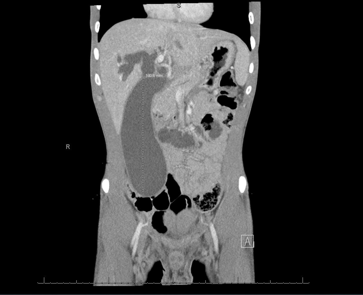
Figure: Coronal CT abdomen revealing a markedly distended gallbladder extending down into the pelvic cavity
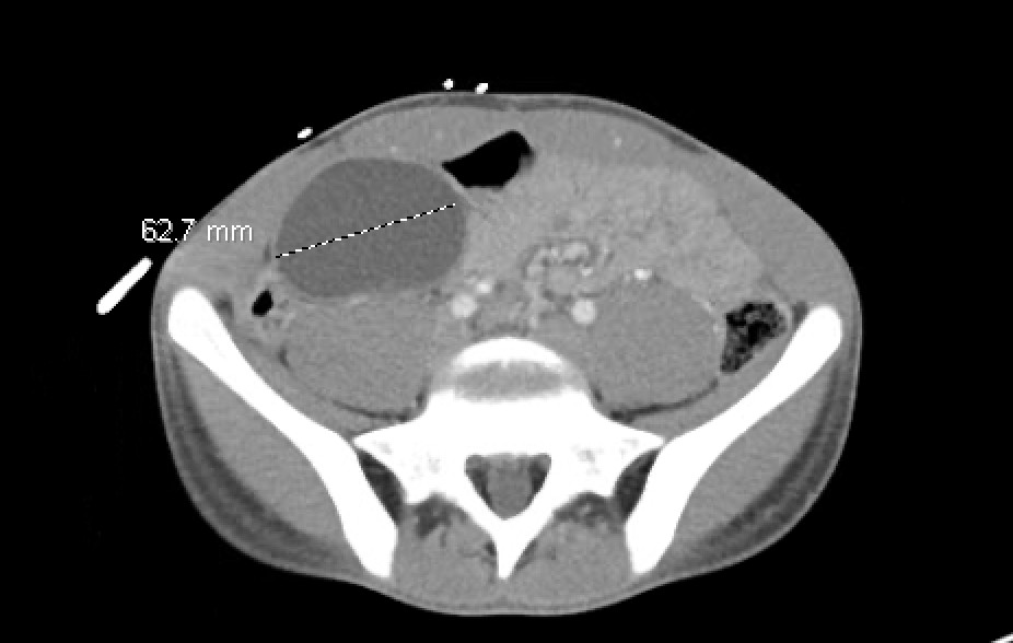
Figure: Axial CT of abdomen
Disclosures: Corey Garcia indicated no relevant financial relationships. Amanda Eukovich indicated no relevant financial relationships. Ivana Rubenstein indicated no relevant financial relationships. Lorraine Chong Tai indicated no relevant financial relationships. Edwin Makarevich indicated no relevant financial relationships. Satya Singh indicated no relevant financial relationships.
Corey Garcia, 1, Amanda Eukovich, DO2, Ivana Rubenstein, DO2, Lorraine Chong Tai, MD2, Edwin Makarevich, DO2, Satya Singh, MD2. P0168 - Mega Gallbladder: A Markedly Distended Gallbladder in a Young Patient With Primary Sclerosing Cholangitis, ACG 2025 Annual Scientific Meeting Abstracts. Phoenix, AZ: American College of Gastroenterology.


 photo")