The Wright Center for Graduate Medical Education Scranton, PA
Seyma Bayram, MD1, Mutaz Kalas, MD2, Ali Osman Avci, MD3, Mehmet Talha Bayram, MD4, Mehmet Bektas, MD5 1The Wright Center for Graduate Medical Education, Scranton, PA; 2Texas Tech University Health Science Center El Paso, El Paso, TX; 3Lokman Hekim University Ankara Hospital, Ankara, Ankara, Turkey; 4Geisinger Wyoming Valley Medical Center, Wilkes-Barre, PA; 5Bayindir Private Hospital, Ankara, Ankara, Turkey Introduction: Annular pancreas, an uncommon congenital anomaly, involves pancreatic tissue encircling the duodenum's second part, with a prevalence estimated at 3-15 cases per 100,000 individuals in various populations. Often asymptomatic and found incidentally in adults, it can manifest with abdominal pain, post-prandial fullness, nausea, or vomiting. This clinical case report aims to explain how annular pancreas can present through recurrent acute pancreatitis episodes, potentially fostering progression to chronic pancreatitis and its associated complications, notably exocrine pancreatic insufficiency.
Case Description/
Methods: A 51-year-old male, with a history of cholecystectomy for a gallbladder polyp, presented with several years of intermittent right upper quadrant abdominal pain. During these episodes, serum amylase and lipase levels were consistently elevated. Contrast-enhanced CT scans during these episodes did not show typical definitive signs of acute pancreatitis. Further investigation with magnetic resonance imaging (MRI) revealed an annular pancreas at the level of the pancreatic head, causing relative narrowing of the second duodenal segment over approximately 3 cm. The pancreatic duct followed a looped course at this particular level, but the common bile duct and main pancreatic duct were otherwise normal. Laboratory tests showed mildly elevated triglycerides and a markedly decreased fecal elastase level, indicative of developing exocrine pancreatic insufficiency. IgG4 levels were normal. Esophagogastroduodenoscopy (EGD) showed a 10 mm subepithelial bulge in the postbulbar duodenum and a broad-based 15 mm subepithelial lesion near the second duodenal portion, adjacent to the annular pancreas identified. Endoscopic ultrasound (EUS) confirmed the annular pancreas and demonstrated a focal, likely benign lesion in the pancreatic head, suggestive of early-stage chronic pancreatitis. Discussion: This case highlights annular pancreas presenting with recurrent episodes of acute pancreatitis and evidence of exocrine pancreatic insufficiency. The diagnosis was established through advanced imaging (MRI, EUS) after initial CT scans were non-diagnostic for typical pancreatitis. This case underscores the importance of considering annular pancreas in the differential diagnosis of recurrent pancreatitis, especially when initial imaging is inconclusive. Comprehensive evaluation, including assessment for exocrine function, is crucial for appropriate management and improving outcomes.
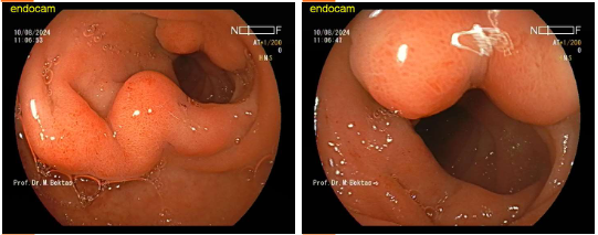
Figure: EGD image showing 10 mm subepithelial bulge in the postbulbar duodenum
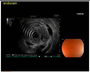
Figure: EUS image showing the pancreatic head circumferentially involving approximately 70% of the duodenal wall.
Disclosures: Seyma Bayram indicated no relevant financial relationships. Mutaz Kalas indicated no relevant financial relationships. Ali Osman Avci indicated no relevant financial relationships. Mehmet Talha Bayram indicated no relevant financial relationships. Mehmet Bektas indicated no relevant financial relationships.
Seyma Bayram, MD1, Mutaz Kalas, MD2, Ali Osman Avci, MD3, Mehmet Talha Bayram, MD4, Mehmet Bektas, MD5. P0106 - Annular Pancreas as a Cause of Recurrent Pancreatitis and Exocrine Insufficiency: A Case Report, ACG 2025 Annual Scientific Meeting Abstracts. Phoenix, AZ: American College of Gastroenterology.