The Wright Center for Graduate Medical Education Scranton, PA
Seyma Bayram, MD1, Alan Jurado, MD2, Mostafa Eysha, MD3, Mehmet Talha Bayram, MD4, Amit Sohagia, MD5 1The Wright Center for Graduate Medical Education, Scranton, PA; 2Department of Internal Medicine at Paul L. Foster School of Medicine, Texas Tech University Health Sciences Center, El Paso TX, USA, El Paso, TX; 3Texas Tech University Health Science Center El Paso, El Paso, TX; 4Geisinger Wyoming Valley Medical Center, Wilkes-Barre, PA; 5Geisinger Community Medical Center, Scranton, PA Introduction: Pancreatic acinar cell carcinoma (ACC) is an uncommon exocrine malignancy, typically affecting older males and presenting with symptoms like abdominal pain or paraneoplastic syndromes. Diagnosis relies on imaging and histopathology, with positive immunostaining for digestive enzymes (e.g., CPA1) being characteristic. While ACC commonly metastasizes to the liver, lungs, and lymph nodes, metastasis to other sites, such as the stomach, is exceptionally rare. Recurrence rates are high, underscoring its aggressive nature. This report details a case of ACC recurring as an isolated gastric intramural mass.
Case Description/
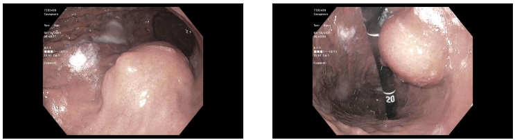
Methods: A 72-year-old male initially presented with abdominal pain and elevated lipase. Imaging revealed a 2.3–2.4 cm lesion in the pancreatic body. Endoscopic ultrasound (EUS) with fine-needle aspiration (FNA) confirmed a poorly differentiated carcinoma, and immunohistochemical staining was consistent with acinar cell carcinoma. The patient subsequently underwent distal pancreatectomy and splenectomy, with surgical pathology revealing grade 2 acinar cell carcinoma (pT2N0) with lymphovascular and perineural invasion. He completed six months of adjuvant modified FOLFIRINOX (mFOLFIRINOX) chemotherapy. Two years later, routine surveillance imaging identified a new 3.7 cm gastric intramural mass near the prior resection site, which was hypermetabolic on PET scan without evidence of distant metastases. Esophagogastroduodenoscopy (EGD) and EUS revealed a submucosal gastric mass, and biopsy confirmed metastatic acinar cell carcinoma with an immunohistochemical profile identical to the original pancreatic tumor. The patient was restarted on mFOLFIRINOX. Discussion: This case illustrates an unusual pattern of metastatic recurrence for pancreatic ACC, presenting as an isolated intramural gastric mass approximately two years after initial resection and adjuvant chemotherapy. Gastric metastasis from pancreatic ACC is exceedingly rare. This highlights the importance of considering atypical metastatic sites in patients with a history of ACC presenting with new gastrointestinal lesions, even after a period of remission. Histopathological confirmation with IHC comparison to the primary tumor is crucial for accurate diagnosis and guiding further management.
Figure: Submucosal Gastric Tumor (3.5 cm in size) in proximal stomach
Disclosures: Seyma Bayram indicated no relevant financial relationships. Alan Jurado indicated no relevant financial relationships. Mostafa Eysha indicated no relevant financial relationships. Mehmet Talha Bayram indicated no relevant financial relationships. Amit Sohagia indicated no relevant financial relationships.
Seyma Bayram, MD1, Alan Jurado, MD2, Mostafa Eysha, MD3, Mehmet Talha Bayram, MD4, Amit Sohagia, MD5. P0105 - Metastatic Recurrence of Pancreatic Acinar Cell Carcinoma to the Stomach, ACG 2025 Annual Scientific Meeting Abstracts. Phoenix, AZ: American College of Gastroenterology.