P0101 - Late Presentation of Metastatic Adenocarcinoma of Bilateral Ovaries, Corpus Uteri, and Right Fallopian Tube From Primary Gallbladder Adenocarcinoma Status Post-Cholecystectomy
Ochsner University Hospital & Clinics Lafayette, LA
Amit Rajkarnikar, MD, Archa Rajesh, DO, Melanie Bienvenu, MD, MPH, Sudhir Aggarwal, MD Ochsner University Hospital & Clinics, Lafayette, LA Introduction: The ovary is a frequent site for metastasis of primary colorectal and gastric tumors. In ovarian malignancies, about 20% of adnexal masses initially suspected to be primary ovarian neoplasms are later identified as metastatic. Malignancies of the gallbladder are rare causes of ovarian tumors and are associated with worse prognosis. We present a case of metastatic adenocarcinoma of bilateral ovaries after s/p cholecystectomy that was found to be gallbladder adenocarcinoma.
Case Description/
Methods: 59-year-old female had acholecystectomy for chronic cholecystitis in 2022. Post surgical histology showed incidental finding of gallbladder adenocarcinoma with lympho-vascular invasion extending to the hepatic margin. CT abdomen/pelvis post-surgery showed mild prominence of the intrahepatic biliary tree and common bile duct and fluid in the gallbladder fossa. She underwent resection of segment 4/5B of the liver, extrahepatic bile duct, complete portal lymphadenectomy, and diagnostic laparoscopy on 01/2023. Segment 4/5 B of liver and portal lymphadenectomy were positive for well differentiated adenocarcinoma consistent with gallbladder origin. Adjuvant chemotherapy was initiated. Bilateral adnexal masses were noted on surveillance CT imaging with interval growth of left adnexal cyst from 4 cm to 5.8 cm and right adnexal cyst from 3.9 to 7.3 cm from 4/2023 to 9/2023. Transvaginal ultrasound showed multiple cervical cysts and multi-septated, and complex bilateral adnexal cysts. Patient underwent total hysterectomy and bilateral salpingo-oophorectomy on 10/2023. Post surgical histology showed metastatic, well-differentiated adenocarcinoma in the left serosal surface of the uterus, the ovaries, and right fallopian tube consistent with GI or biliary origin. Tumoral cells were positive for CK7, CK20, CEA, CDX2, with a higher proliferation rate by Ki-67. In view of clinical history, the findings were consistent with metastasis from a gallbladder adenocarcinoma. Discussion: Differentiating between primary and metastatic tumors poses significant diagnostic challenges for both clinicians and pathologists. The exact pathway for gastrointestinal metastases to the ovary remains unclear. Evidence from previous research on metastatic ovarian cancer of colonic origin suggests that most cases involve metastases to the ovarian stroma. Accurate differentiation and distinction between primary and metastatic tumors are essential. Appropriate testing should be utilized as a guide to aid in appropriate treatment strategies.
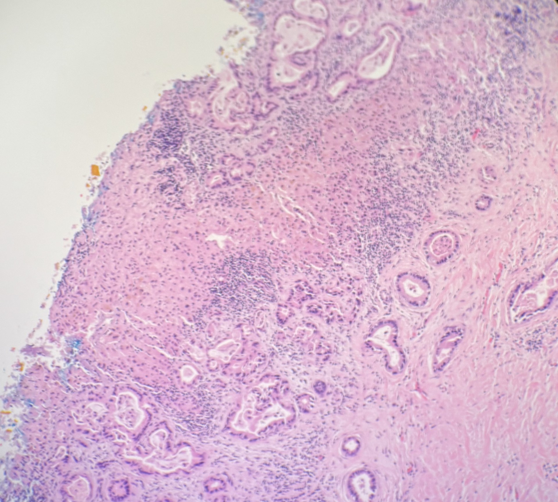
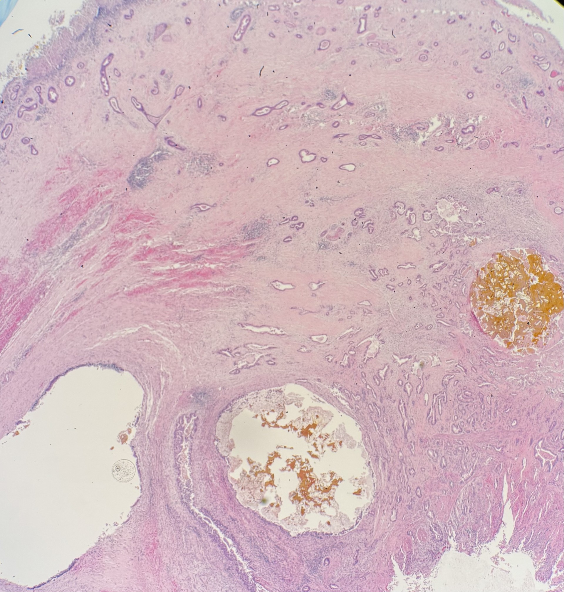
Figure: H&E stain showing lumen to liver margin with adenocarcinoma.
Disclosures: Amit Rajkarnikar indicated no relevant financial relationships. Archa Rajesh indicated no relevant financial relationships. Melanie Bienvenu indicated no relevant financial relationships. Sudhir Aggarwal indicated no relevant financial relationships.
Amit Rajkarnikar, MD, Archa Rajesh, DO, Melanie Bienvenu, MD, MPH, Sudhir Aggarwal, MD. P0101 - Late Presentation of Metastatic Adenocarcinoma of Bilateral Ovaries, Corpus Uteri, and Right Fallopian Tube From Primary Gallbladder Adenocarcinoma Status Post-Cholecystectomy, ACG 2025 Annual Scientific Meeting Abstracts. Phoenix, AZ: American College of Gastroenterology.

 photo")